

Vascular calcification: pathophysiology and risk factors
- PMID: 22476974
- PMCID: PMC3959826
- DOI: 10.1007/s11906-012-0265-8
Vascular calcification: pathophysiology and risk factors
Abstract
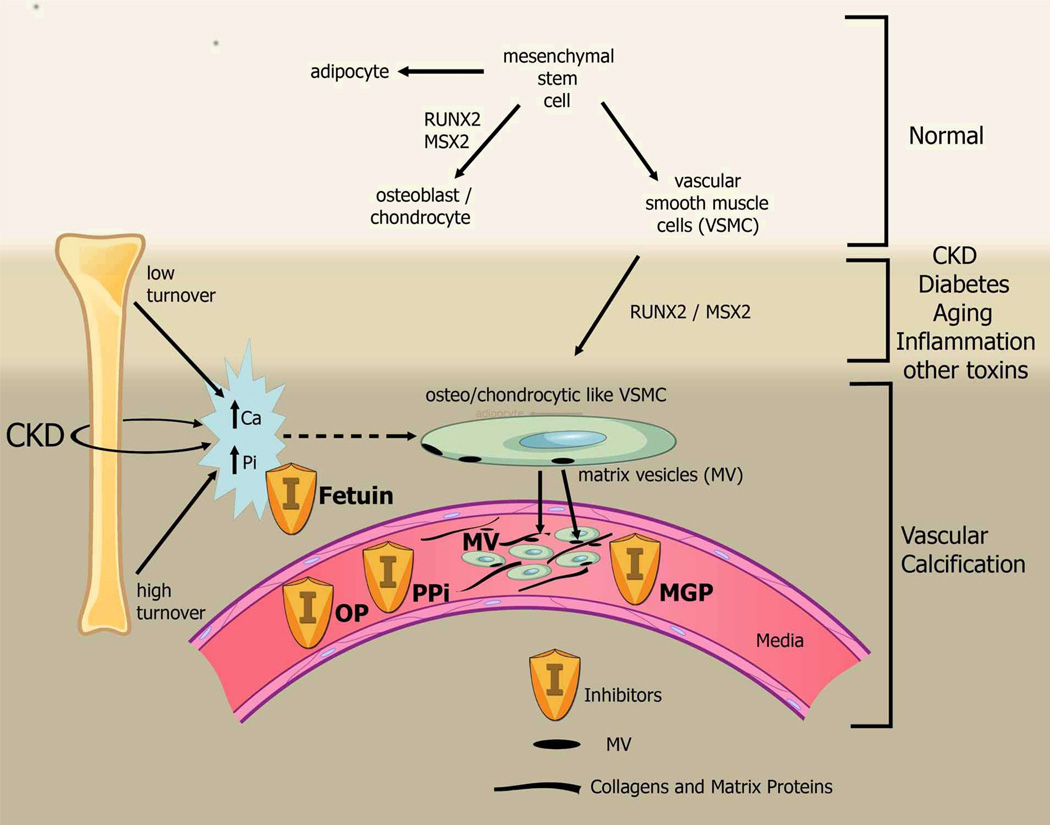
Vascular calcification can occur in nearly all arterial beds and in both the medial and intimal layers. The initiating factors and clinical consequences depend on the underlying disease state and location of the calcification. The best studied manifestation is coronary artery calcification, in part because of the obvious clinical consequences, but also because of CT-based imaging modalities. In the general population, the presence of coronary artery calcification increases cardiovascular risk above that predicted by traditional Framingham risk factors, suggesting the presence of nontraditional risk factors. In patients with chronic kidney disease (CKD), coronary artery calcification is more prevalent and markedly more severe than in the general population. In these CKD patients, nontraditional risk factors such as oxidative stress, advanced glycation end products, and disordered mineral metabolism are also more prevalent and more severe and offer mechanistic insight into the pathogenesis of vascular calcification.
Figures
References
-
- Moe SM, Chen NX. Mechanisms of vascular calcification in chronic kidney disease. J Am Soc Nephrol. 2008;19(2):213–216. - PubMed
-
- Ibanez B, Badimon JJ, Garcia MJ. Diagnosis of atherosclerosis by imaging. Am J Med. 2009;122(1 Suppl):S15–S25. - PubMed
-
- Moe SM, Chen NX. Pathophysiology of vascular calcification in chronic kidney disease. Circ Res. 2004;95(6):560–567. - PubMed
-
- Proudfoot D, Shanahan CM, Weissberg PL. Vascular calcification: new insights into an old problem [editorial; comment] Journal of Pathology. 1998;185(1):1–3. - PubMed
-
- Lehto S, Niskanen L, Suhonen M, Ronnemaa T, Laakso M. Medial artery calcification. A neglected harbinger of cardiovascular complications in non-insulin-dependent diabetes mellitus. Arterioscler Thromb Vasc Biol. 1996;16(8):978–983. - PubMed
Publication types
MeSH terms
Substances
Grants and funding
LinkOut - more resources
Full Text Sources
Other Literature Sources
Medical
