

doi: 10.3349/ymj.2012.53.3.654.
Phagocytosis and endocytosis of silver nanoparticles induce interleukin-8 production in human macrophages
Affiliations
- PMID: 22477013
- PMCID: PMC3343436
- DOI: 10.3349/ymj.2012.53.3.654
Item in Clipboard
Phagocytosis and endocytosis of silver nanoparticles induce interleukin-8 production in human macrophages
Yonsei Med J.
2012 May.
Abstract
Phagocytosis or endocytosis by macrophages is critical to the uptake of fine particles, including nanoparticles, in order to initiate toxic effects in cells. Here, our data enhance the understanding of the process of internalization of silver nanoparticles by macrophages. When macrophages were pre-treated with inhibitors to phagocytosis, caveolin-mediated endocytosis, or clathrin-mediated endocytosis, prior to exposure to silver nanoparticles, Interleukin-8 (IL-8) production was inhibited. Although cell death was not reduced, the inflammatory response by macrophages was compromised by phagocytosis and endocytosis inhibitors.
Conflict of interest statement
The authors have no financial conflicts of interest.
Figures
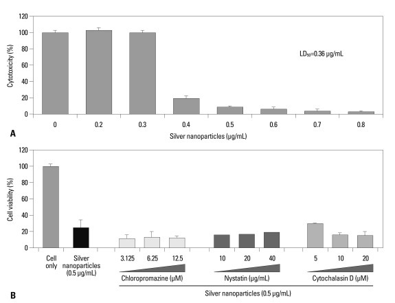
Cytotoxicity of silver nanoparticles in macrophages. (A) The cytotoxicity in U937 cells were assessed by CCK-8 assay. The LD50 of 5-nm silver nanoparticles was 0.36 µg/mL. (B) Each inhibitors were treaetd 1 hour before exposure to 0.5 µg/mL of 5-nm silver nanoparticles.
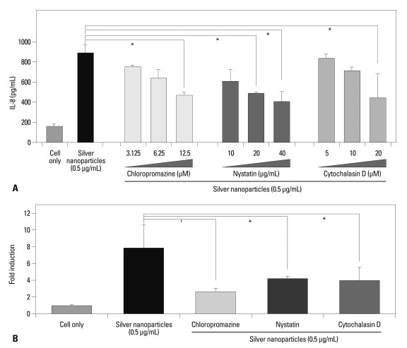
Effects of inhibitors on IL-8 production induced by silver nanoparticles. Each inhibitors were added 1 hour before treatment of nanoparticles. (A) IL-8 in cell culture supernatants were assessed by ELISA 18 hours after exposure to 5-nm silver nanoparticles. (B) Real-time RT-PCR was performent. RNA was perpared from cells treated with 5-nm silver nanoparticles for 2 hours. Chloropromazine was treated at 12.5 µm, nystatin, at 40 µg/mL and cytochalasin D, at 20 µM. *p<0.05, †p<0.001.
Similar articles
-
The effects of sub-lethal concentrations of silver nanoparticles on inflammatory and stress genes in human macrophages using cDNA microarray analysis.Biomaterials. 2012 Jun;33(18):4690-9. doi: 10.1016/j.biomaterials.2012.03.006. Epub 2012 Mar 27. Biomaterials. 2012. PMID: 22459196
-
Size dependent macrophage responses and toxicological effects of Ag nanoparticles.Chem Commun (Camb). 2011 Apr 21;47(15):4382-4. doi: 10.1039/c1cc10357a. Epub 2011 Mar 10. Chem Commun (Camb). 2011. PMID: 21390403
-
A novel type of silver nanoparticles and their advantages in toxicity testing in cell culture systems.Arch Toxicol. 2012 Jul;86(7):1089-98. doi: 10.1007/s00204-012-0836-0. Epub 2012 Mar 29. Arch Toxicol. 2012. PMID: 22456835
-
Cellular uptake, intracellular trafficking and cytotoxicity of silver nanoparticles.Toxicol Lett. 2012 Sep 3;213(2):249-59. doi: 10.1016/j.toxlet.2012.07.009. Epub 2012 Jul 20. Toxicol Lett. 2012. PMID: 22820426
-
Silver Nanoparticle-Mediated Cellular Responses in Various Cell Lines: An in Vitro Model.Int J Mol Sci. 2016 Sep 22;17(10):1603. doi: 10.3390/ijms17101603. Int J Mol Sci. 2016. PMID: 27669221 Free PMC article. Review.
Cited by
-
Unlocking the Potential of Silver Nanoparticles: From Synthesis to Versatile Bio-Applications.Pharmaceutics. 2024 Sep 21;16(9):1232. doi: 10.3390/pharmaceutics16091232. Pharmaceutics. 2024. PMID: 39339268 Free PMC article. Review.
-
Pro-inflammatory effects of silver nanoparticles in the intestine.Arch Toxicol. 2022 Jun;96(6):1551-1571. doi: 10.1007/s00204-022-03270-w. Epub 2022 Mar 16. Arch Toxicol. 2022. PMID: 35296919 Review.
-
Metal Organic Framework (MOF) Particles as Potential Bacteria-Mimicking Delivery Systems for Infectious Diseases: Characterization and Cellular Internalization in Alveolar Macrophages.Pharm Res. 2019 Feb 21;36(4):53. doi: 10.1007/s11095-019-2589-4. Pharm Res. 2019. PMID: 30790066
-
Food additives can act as triggering factors in celiac disease: Current knowledge based on a critical review of the literature.World J Clin Cases. 2019 Apr 26;7(8):917-927. doi: 10.12998/wjcc.v7.i8.917. World J Clin Cases. 2019. PMID: 31119137 Free PMC article. Review.
-
Implementation of a Dynamic Co-Culture Model Abated Silver Nanoparticle Interactions and Nanotoxicological Outcomes In Vitro.Nanomaterials (Basel). 2021 Jul 12;11(7):1807. doi: 10.3390/nano11071807. Nanomaterials (Basel). 2021. PMID: 34361193 Free PMC article.
References
-
- Hackenberg S, Scherzed A, Kessler M, Hummel S, Technau A, Froelich K, et al. Silver nanoparticles: evaluation of DNA damage, toxicity and functional impairment in human mesenchymal stem cells. Toxicol Lett. 2011;201:27–33. - PubMed
-
- Tantra R, Knight A. Cellular uptake and intracellular fate of engineered nanoparticles: a review on the application of imaging techniques. Nanotoxicology. 2011;5:381–392. - PubMed
-
- Zhao F, Zhao Y, Liu Y, Chang X, Chen C, Zhao Y. Cellular uptake, intracellular trafficking, and cytotoxicity of nanomaterials. Small. 2011;7:1322–1337. - PubMed
Publication types
MeSH terms
Substances
LinkOut - more resources
Full Text Sources
