Review
. 2012 Apr;95(4):333-45.
doi: 10.1007/s12185-012-1059-0. Epub 2012 Apr 5.

Protein C anticoagulant and cytoprotective pathways

Affiliations
Review

Protein C anticoagulant and cytoprotective pathways

John H Griffin et al. Int J Hematol. 2012 Apr.

Abstract

Plasma protein C is a serine protease zymogen that is transformed into the active, trypsin-like protease, activated protein C (APC), which can exert multiple activities. For its anticoagulant action, APC causes inactivation of the procoagulant cofactors, factors Va and VIIIa, by limited proteolysis, and APC's anticoagulant activity is promoted by protein S, various lipids, high-density lipoprotein, and factor V. Hereditary heterozygous deficiency of protein C or protein S is linked to moderately increased risk for venous thrombosis, while a severe or total deficiency of either protein is linked to neonatal purpura fulminans. In recent years, the beneficial direct effects of APC on cells which are mediated by several specific receptors have become the focus of much attention. APC-induced signaling can promote multiple cytoprotective actions which can minimize injuries in various preclinical animal injury models. Remarkably, pharmacologic therapy using APC demonstrates substantial neuroprotective effects in various murine injury models, including ischemic stroke. This review summarizes the molecules that are central to the protein C pathways, the relationship of pathway deficiencies to venous thrombosis risk, and mechanisms for the beneficial effects of APC.


Figures

Figure 1
Figure 1. Schematic models of Protein C activation and expression of APC activities
(a) Physiologic activation of protein C (PC) by the thrombin (IIa)-thrombomodulin (TM) complex occurs on the surface of endothelial cells. IIa bound to TM activates PC, especially when PC is bound to the endothelial receptor (EPCR). Since protein C and APC have a similar affinity for EPCR, after activation APC can either dissociate from EPCR to exert anticoagulant activity or remain bound to EPCR where it might express multiple direct cellular activities (b). Dissociation of APC from EPCR allows expression of APC’s anticoagulant activity on cell membrane surfaces, various microparticles, or lipoproteins (e.g., High Density Lipoprotein). As an anticoagulant, APC cleaves the activated cofactors Va (fVa) and VIIIa (fVIIIa) to yield inactivated cofactors, fVi and fVIIIi. This proteolytic inactivation is enhanced by protein cofactors (e.g., protein S, factor V) and lipids cofactors (e.g., phosphatidylserine, cardiolipin, glucosylceramide, or HDL). (b) PAR1 mediates multiple cytoprotective effects of APC. In most, but not all, reported studies of APC’s beneficial effects of on endothelial cells, the cellular receptors EPCR and PAR1 are required. These cytoprotective effects include anti-apoptotic activities, anti-inflammatory activities, protection of endothelial barrier functions, and favorable alteration of gene expression profiles. This paradigm in which EPCR-bound APC activates PAR1 to initiate signaling is consistent with many in vitro and in vivo data. Localization of APC signaling in the caveolin-1 rich microdomains (caveolae) may help differentiate mechanisms for cytoprotective APC signaling versus proinflammatory thrombin signaling. Additional mechanisms for APC effects on cells may involve other receptors. These effects include APC anti-inflammatory effects on leukocytes or cytoprotective effects on dendritic cells and neurons. Other receptors may include PAR3, various integrins (e.g., Mac-1 (CD11b/CD18), β1 integrins, β3 integrins), S1P1, or apolipoprotein E receptor 2 (LRP8). This scheme is taken from Blood. (LO Mosnier et al. The cytoprotective protein C pathway. Blood 2007 109:3161–3172. © the American Society of Hematology)
Figure 2
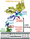
Figure 2. Protein S polypeptide scheme showing multiple domains and protein S Tokushima mutation
Specific protein S domains are color coded and labeled. The N-terminal cluster of domains is responsible for binding to APC. The amino acid side chain of residue 155 is shown in red, representing the protein S Tokushima polymorphism, K155E (“155” is the mature protein S numbering which equates to K196E in full length precursor numbering). The schematic model of protein S was based on available models and structures of the individual GLA-TSR-EGF1 [167], EGF3–4 (1Z6C) [168], and the SHBG [169] domains. EGF2 was modeled by homology modeling using Swiss Model based on templates for extracellular domain of the LDL receptor (1N7DA), low-density lipoprotein receptor (1HZ8A), low-density lipoprotein receptor (1HJ7A) and EGF-like module-containing mucin-like hormone receptor-like 2 (2BO2A) as templates [170]. The individual domains were put together using Modeller [171], displayed and colored in Accelrys Discovery Studio and rendered in POV-Ray.
Figure 3
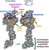
Figure 3. APC space-filling model showing mutations that differentiate amino acid residue requirements for anticoagulant versus cytoprotective activities
The model of APC extending from bottom to top depicts the N-terminal Gla domain at the bottom which binds EPCR and phospholipids membranes. The protease domain is at at the top, with the EGF-1and EGF-2 domains in the middle section of the model. The "active site" triad of Ser, His and Asp residues is noted in red. Green highlights the L38D mutation that reduces anticoagulant activity due to reduced protein S enhancement. Gold highlights mutations of E330 and E333 to Ala that selectively reduce PAR1 signaling and the E149A mutation in the C-terminus of the light chain that causes loss of cytoprotective anti-inflammatory and anti-apoptotic activities but gain-of-function of anticoagulant activity due to enhanced protein S cofactor effects. On the top of the model, blue highlights five basic residues (KKK191–193 and RR229/230) which form a large positively charged exosite that recognizes factor Va. When these five basic residues are mutated to Ala, < 10 % anticoagulant activity remains whereas cytoprotective activities remain intact. Purple highlights two residues (R222 and D237 that are not in view) which when mutated to Cys can form a disulfide bond, causing loss of most anticoagulant activity but retention of cytoprotective activity. The model of full length APC [172] is based on the serine protease domain structure of APC (Protein Data Bank entry 1AUT [155]).
Figure 4
Figure 4. APC’s neuroprotective actions on the neurovascular unit following ischemic injury
Ischemia promotes endothelial cell apoptosis, breakdown of the blood-brain-barrier (BBB), neuroinflammation, and damage to neurons. By its cytoprotective actions, APC protects vascular integrity and ameliorates post-ischemic BBB breakdown, thereby preventing secondary neuronal damage mediated by entry of several blood-derived neurotoxic and vasculotoxic molecules. APC can cross an intact BBB via EPCR-dependent transport to reach its neuronal targets in brain. APC can express direct neuronal protective activity to prevent neuron damage. APC also expresses anti-inflammatory activities by blocking early post-ischemic infiltration of brain by neutrophils and by suppressing microglia activation.

Similar articles

Cited by

References

    1. Esmon CT. Inflammation and the activated protein C anticoagulant pathway. Semin.Thromb.Hemost. 2006;32:49–60. - PubMed
    1. Rezaie AR. Regulation of the protein C anticoagulant and antiinflammatory pathways. Curr.Med.Chem. 2010;17:2059–2069. - PMC - PubMed
    1. Riewald M, Ruf W. Science review: Role of coagulation protease cascades in sepsis. Crit.Care. 2003;7:123–129. - PMC - PubMed
    1. Weiler H, Kerschen E. Modulation of sepsis outcome with variants of activated protein C. J Thromb Haemost. 2009;7:127–131. - PubMed
    1. Jackson CJ, Xue M. Activated protein C--an anticoagulant that does more than stop clots. Int.J.Biochem.Cell Biol. 2008;40:2692–2697. - PubMed