

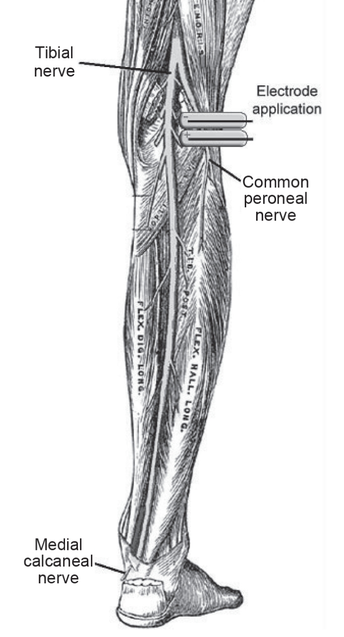
Augmentation of venous, arterial and microvascular blood supply in the leg by isometric neuromuscular stimulation via the peroneal nerve
- PMID: 22477572
- PMCID: PMC2949997
- DOI: 10.1055/s-0031-1278361
Augmentation of venous, arterial and microvascular blood supply in the leg by isometric neuromuscular stimulation via the peroneal nerve
Abstract
Background: Deep vein thrombosis (DVT) is the formation of a blood clot within the deep veins. During periods of sitting, blood flow is decreased and this contributes to an increased risk of DVT. Trials have shown that 5% to 10% of passengers undertaking long-haul flights develop asymptomatic calf DVT.
Aim: To investigate the safety and efficacy of a novel neuromuscular device that augments peripheral blood flow.
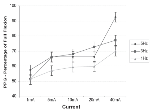
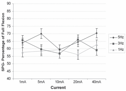
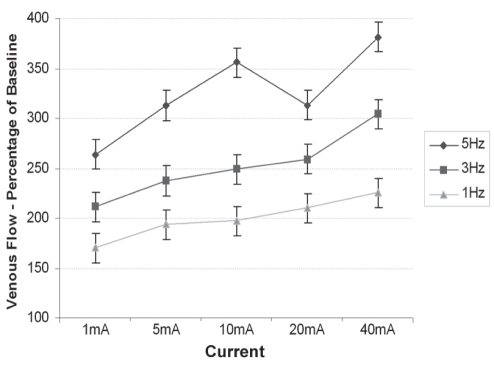
Methods: Thirty healthy volunteers were assessed while seated. Each subject had one leg connected to the stimulator and the other leg immobile acting as control. Fifteen sequential electrical stimulations were applied for 5 min each followed by a 10 min recovery phase. The following noninvasive measurements were performed before, during and after the stimulation programs: photoplethysmography, strain gauge plethysmography, laser Doppler fluxmetry, transcutaneous oxygen tension, pulse oximetry, superficial femoral vein blood flow and vessel diameter (ultrasound); discomfort questionnaires were also administered.
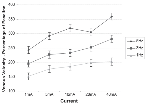
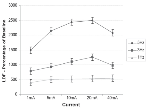
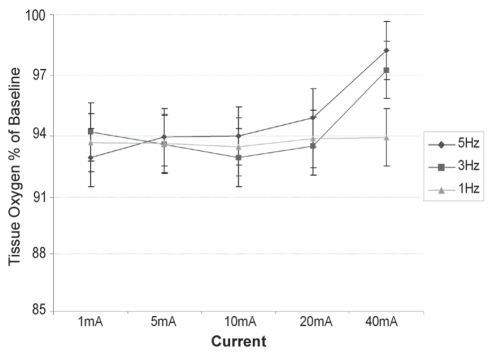
Results: During neuromuscular stimulation, significant increases in blood volume flow and velocity and skin capillary blood flow were found; transdermal skin oxygen levels were maintained. No changes were observed in heart rate, blood pressure, oxygen saturation or femoral vein vessel diameter.
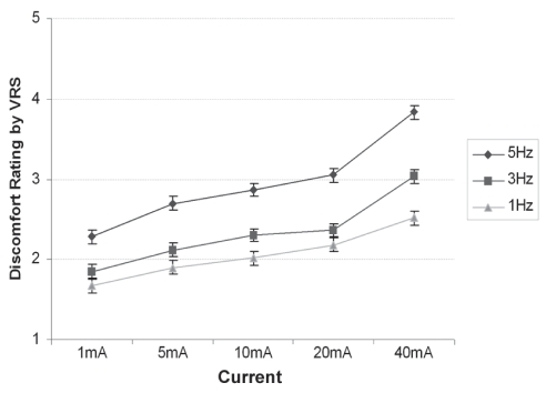
Conclusions: Using a newly developed device, electrical nerve stimulation of the lower leg significantly increased blood flow; the device in the present study is, therefore, a promising tool for the development of a novel DVT prevention device. Because this method of electrical nerve stimulation is virtually pain free, the present study has significant implications for the prevention of DVT in hospitals, outpatient settings and community care settings, as well as in preventing travel-related thrombosis.
Figures
Similar articles
-
Measurement of blood flow in the deep veins of the lower limb using the geko™ neuromuscular electro-stimulation device.Int Angiol. 2016 Aug;35(4):406-10. Epub 2016 Mar 2. Int Angiol. 2016. PMID: 26934561 Clinical Trial.
-
Haemodynamic changes with the use of neuromuscular electrical stimulation compared to intermittent pneumatic compression.Phlebology. 2015 Jun;30(5):365-72. doi: 10.1177/0268355514531255. Epub 2014 Apr 10. Phlebology. 2015. PMID: 24722790 Clinical Trial.
-
Endovascular laser therapy for varicose veins: an evidence-based analysis.Ont Health Technol Assess Ser. 2010;10(6):1-92. Epub 2010 Apr 1. Ont Health Technol Assess Ser. 2010. PMID: 23074409 Free PMC article.
-
Diagnostic imaging in deep vein thrombosis of the limbs.Rays. 1996 Jul-Sep;21(3):328-39. Rays. 1996. PMID: 9063053 Review. English, Italian.
-
Pathophysiology and diagnosis of deep venous thrombosis.Semin Nucl Med. 2001 Apr;31(2):90-101. doi: 10.1053/snuc.2001.21406. Semin Nucl Med. 2001. PMID: 11330789 Review.
Cited by
-
Effects on venous flow of transcutaneous electrical stimulation, neuromuscular stimulation, and sham stimulation on soleus muscle: A randomized crossover study in healthy subjects.Medicine (Baltimore). 2022 Sep 2;101(35):e30121. doi: 10.1097/MD.0000000000030121. Medicine (Baltimore). 2022. PMID: 36107611 Free PMC article. Clinical Trial.
-
Design of a Device for Lower Limb Prophylaxis and Exercise.IEEE J Transl Eng Health Med. 2020 Nov 9;9:2100107. doi: 10.1109/JTEHM.2020.3037018. eCollection 2021. IEEE J Transl Eng Health Med. 2020. PMID: 33224639 Free PMC article.
-
The impact of a new intervention for venous leg ulcers: A within-patient controlled trial.Int Wound J. 2023 Aug;20(6):2260-2268. doi: 10.1111/iwj.14107. Epub 2023 Feb 13. Int Wound J. 2023. PMID: 36785909 Free PMC article.
-
A feasibility randomised controlled trial to evaluate the effectiveness of a novel neuromuscular electro-stimulation device in preventing the formation of oedema following total hip replacement surgery.Heliyon. 2018 Jul 18;4(7):e00697. doi: 10.1016/j.heliyon.2018.e00697. eCollection 2018 Jul. Heliyon. 2018. PMID: 30094367 Free PMC article.
-
Clinical Study of Neuromuscular Electrical Stimulation in the Prevention of Deep Venous Thrombosis of Lower Extremities after Anterior Cruciate Ligament Reconstruction.J Healthc Eng. 2022 Mar 11;2022:7857272. doi: 10.1155/2022/7857272. eCollection 2022. J Healthc Eng. 2022. Retraction in: J Healthc Eng. 2023 Aug 30;2023:9856721. doi: 10.1155/2023/9856721. PMID: 35310181 Free PMC article. Retracted. Clinical Trial.
References
-
- Malone PC, Agutter PS. The aetiology of deep venous thrombosis. QJM. 2006;99:581–93. - PubMed
-
- Sevitt S. Pathology and pathogenesis of deep vein thrombosis. In: Poller L, editor. Recent Advances in Thrombosis. Edinburgh: Churchill Livingstone; 1973. pp. 17–38.
-
- Nicolaides AN, Kakkar VV, Field ES, Renney JT. The origin of deep vein thrombosis: A venographic study. Br J Radiol. 1971;44:653–63. - PubMed
-
- Nicolaides AN, Breddin HK, Fareed J, et al. Prevention of venous thromboembolism. International Consensus Statement. Guidelines compiled in accordance with the scientific evidence. Int Angiol. 2001;20:1–37. - PubMed
-
- Kim YH. The incidence of deep vein thrombosis after cementless and cemented knee replacement. J Bone Joint Surg Br. 1990;72:779–83. - PubMed
LinkOut - more resources
Full Text Sources
Other Literature Sources
Medical
