. 2012;8(3):e1002566.
doi: 10.1371/journal.pgen.1002566. Epub 2012 Mar 29.

Mammalian neurogenesis requires Treacle-Plk1 for precise control of spindle orientation, mitotic progression, and maintenance of neural progenitor cells

Affiliations

Mammalian neurogenesis requires Treacle-Plk1 for precise control of spindle orientation, mitotic progression, and maintenance of neural progenitor cells

Daisuke Sakai et al. PLoS Genet. 2012.

Abstract

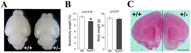
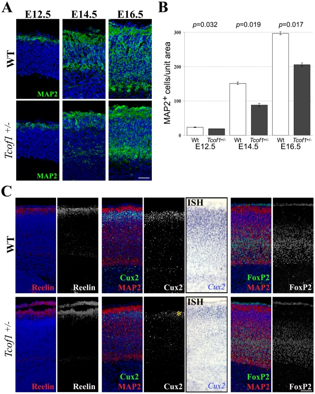
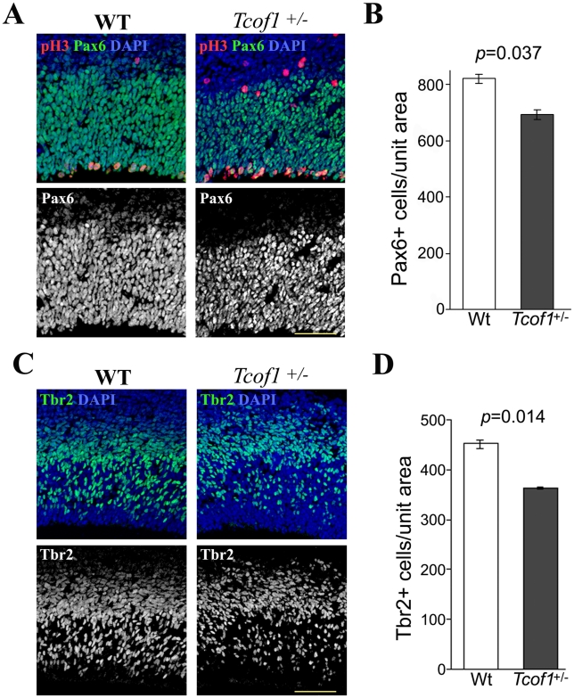
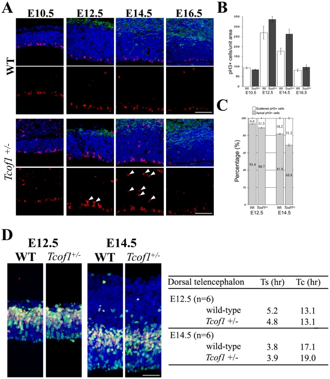
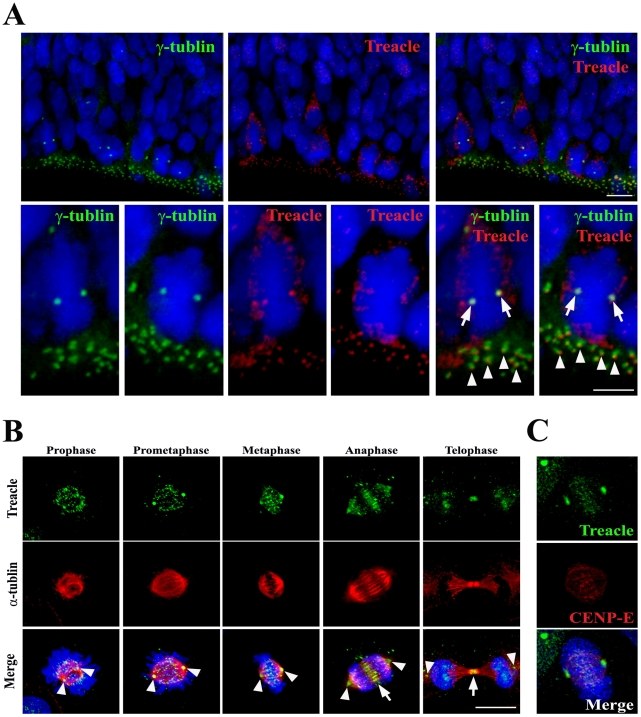
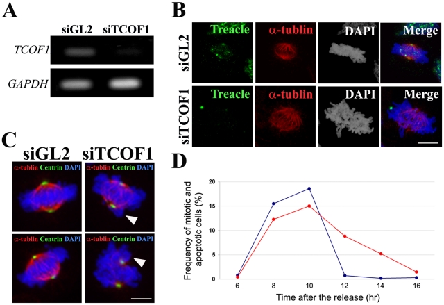
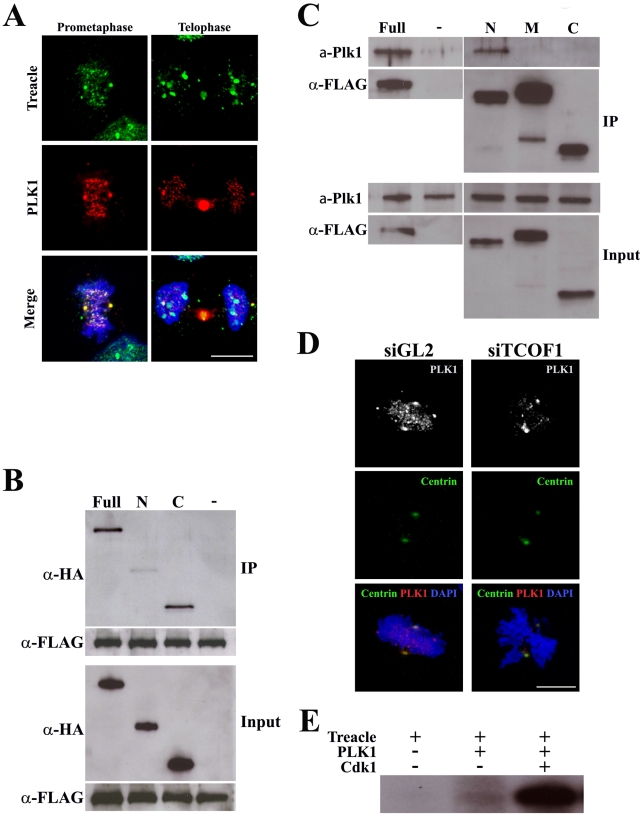
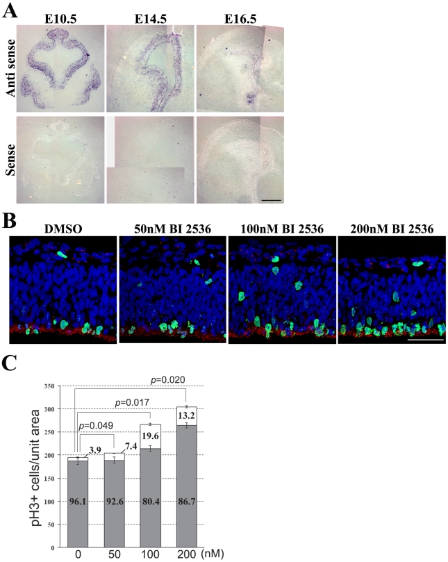
The cerebral cortex is a specialized region of the brain that processes cognitive, motor, somatosensory, auditory, and visual functions. Its characteristic architecture and size is dependent upon the number of neurons generated during embryogenesis and has been postulated to be governed by symmetric versus asymmetric cell divisions, which mediate the balance between progenitor cell maintenance and neuron differentiation, respectively. The mechanistic importance of spindle orientation remains controversial, hence there is considerable interest in understanding how neural progenitor cell mitosis is controlled during neurogenesis. We discovered that Treacle, which is encoded by the Tcof1 gene, is a novel centrosome- and kinetochore-associated protein that is critical for spindle fidelity and mitotic progression. Tcof1/Treacle loss-of-function disrupts spindle orientation and cell cycle progression, which perturbs the maintenance, proliferation, and localization of neural progenitors during cortical neurogenesis. Consistent with this, Tcof1(+/-) mice exhibit reduced brain size as a consequence of defects in neural progenitor maintenance. We determined that Treacle elicits its effect via a direct interaction with Polo-like kinase1 (Plk1), and furthermore we discovered novel in vivo roles for Plk1 in governing mitotic progression and spindle orientation in the developing mammalian cortex. Increased asymmetric cell division, however, did not promote increased neuronal differentiation. Collectively our research has therefore identified Treacle and Plk1 as novel in vivo regulators of spindle fidelity, mitotic progression, and proliferation in the maintenance and localization of neural progenitor cells. Together, Treacle and Plk1 are critically required for proper cortical neurogenesis, which has important implications in the regulation of mammalian brain size and the pathogenesis of congenital neurodevelopmental disorders such as microcephaly.


Conflict of interest statement

The authors have declared that no competing interests exist.

Figures

Figure 1
Figure 1. Tcof1 heterozygous mutant mice show small brain.
(A) Dorsal view of wild-type (+/+) and Tcof1 heterozygous mutant (+/−) brains of P14 mice. (B) Brain weight per body weight and body weight of wild-type (n = 6) and Tcof1 +/− mice (n = 6). (C) Coronal sections of wild-type (+/+) and Tcof1 heterozygous (+/−) cerebrum stained with HE. The brain is much smaller, but tissue architecture and morphology is normal. Scale Bars: C, 200 µm.
Figure 2
Figure 2. Abnormal brain development resulting from Treacle deficiency.
(A) Neurons in E12.5–16.5 embryonic forebrains were visualized by immunofluorescence with anti-MAP2 antibody (green). (B) Quantification of neurons in forebrain. MAP2-positive cells in the wild-type and Tcof1 +/− brain were counted in a unit section of 100 µm width. (C) Immunostaining of cortical layers with anti-Reelin (layer I), anti-Cux2 (layer II, III and IV; asterisk) and anti-FoxP2 (layer V and VI) antibodies and in situ hybridization for Cux2 on coronal sections of E18.5 wild-type and Tcof1 +/− mice. To observe the cortical cortex, MAP2-positive neurons (red) are co-stained with Cux2 and FoxP2 (green). Scale Bars: A, 25 µm; C, 100 µm.
Figure 3
Figure 3. Tcof1 deficiency affects the number of Pax6-positive apical progenitors and Tbr2-positive basal progenitor cells in the telencephalon.
(A) Immunofluorescence detection of Pax6-positive progenitor cells (green) and anti-phospho-Histone H3 antibody (red) mitotic cells in the telencephalon of E14.5 wild-type and Tcof1 +/− embryos. Tissue sections were counterstained with DAPI (blue). (B) Bar graph depicting the number of Pax6-positive cell in the wild-type and Tcof1 +/− brain in a unit section of 125 µm width. (C) Co-immunostaining of the neuroepithelium of E14.5 wild-type and Tcof1 +/− embryos for Tbr2-positive neurons (green) and pH 3-positive (red) mitotic cells (D). Bar graph quantifying the number of Tbr2-positive cells in Tcof1 +/− embryos and their wild-type littermates in a unit section of 100 µm width. Scale Bars: A and C, 20 µm.
Figure 4
Figure 4. Neural progenitor cells in Tcof1 +/− embryos exhibit mitotic defects.
(A) Immunostaining of E10.5–E16.5 embryonic forebrains using a pH 3 antibody (red). Arrowheads indicate scattered progenitor cells in the ventricular and subventricular zones of the telencephalon in E14.5 Tcof1 +/− embryos. The neuron layers were visualized by immunofluorescence of anti-MAP2 antibody (green). (B) Bar graph depicting average numbers of mitotic cells in the telencephalon of E10.5–E16.5 wild-type and Tcof1 +/− embryos, counted in a unit section of 100 µm width. (C) Bar graph depicting the percentage of apical neural progenitor cells and abnormal scattered neural progenitor cells in the telencephalon of E12.5 and E14.5 wild-type and Tcof1 +/− embryos. (D) Co-labeling of the telencehalon in wild-type and Tcof1 mutant embryos with IdU (green) and BrdU (red). S-phase and total cell cycle length were estimated by the number of IdU- and BrdU-positive cells and revealed an increase in total cell cycle length in Tcof1 +/− embryos compared to wild-type littermates. Scale Bars: A and D, 50 µm.
Figure 5
Figure 5. Dynamic localization of Treacle in mitotic cells.
(A) Immunofluorescence images of wild-type forebrain at E11.5 detecting Treacle (red), the centrosome (γ-tubulin; green) and nuclei (DAPI; blue). Arrowheads and arrows indicate Treacle localization at the centrosome in interphase cells and mitotic cells respectively. (B) Immunofluorescence images of HeLa cells showing the dynamic localization of Treacle (green) during mitosis, particularly at the centrosomes (arrowheads) and kinetochore in prophase, prometaphase and metaphase cells as well as at the midzone during anaphase (arrow) and at the midbody (arrow) in telophase. The microtubular networks are detected by immunostaining with an anti-α-tubulin antibody. (C) Treacle co-localizes with the kinetochore marker, CENP-E. Scale Bars: A and B, 10 µm.
Figure 6
Figure 6. Essential function of Treacle in mitotic spindle formation and mitotic progression.
(A) PCR analysis of efficacy of TCOF1 knockdown in HeLa cells 48 hours after control (siGL2) or TCOF1 (si TCOF1) siRNA transfection. (B) Immunostaining of mitotic HeLa cells in control knock-down (siGL2) and TCOF1 knock-down (si TCOF1) cultures analyzed with anti-α-tubulin (red) and anti-Treacle (green) antibodies. (C) Mitotic cells in TCOF1knock-down (siTCOF1) and control (siGL2) cultures immunostained with anti-α-tubulin (red) and anti-centrin (green) antibodies. Arrowheads indicate abnormal mitotic spindle and chromosome alignment in TCOF1 knock-down cultures. (D) Graph depicting the frequency of mitotic cells labeled via immunostaining with a pH 3 antibody at 6–16 hours post siGL2 or siTCOF1 transfection and the marked delay in mitotic exit exhibited by TCOF1 knock-down cells. Scale Bars: B and C, 5 µm.
Figure 7
Figure 7. Treacle interacts with PLK1 and mediates its localization.
(A) Immunofluorescent localization of Treacle (green) and PLK1 (red) in mitotic HeLa cells. (B) Interaction between FLAG-tagged Treacle and HA-tagged Plk1 as evidenced by immunoprecipitation (IP) using an anti-FLAG antibody. Precipitated Plk1 and Treacle proteins were detected by western analysis using anti-HA and anti-FLAG antibodies, respectively. Full-length (lane Full) and C-terminal half (lane C) of Plk1 are detected in the immunoprecipitated fraction. PLK1 is not detected in the immunoprecipitated fraction of untransfected HeLa cells (lane -). (C) Interaction between FLAG-tagged Treacle and endogenous PLK1 was examined by IP using anti-FLAG antibody. Precipitated PLK1 and Treacle proteins were detected by western analysis using anti-PLK1 and anti-FLAG antibodies, respectively. Endogenous PLK1 binds to FLAG-tagged full length (lane Full) and N-terminal part (lane N) of Treacle in HeLa cells. PLK1 is not detected in the immunoprecipitated fraction of untransfected HeLa cells (lane -). (D) HeLa cells immunolabelled for PLK1 (white and red) and centrin (green) 24 hours after control (siGL2) or Tcof1 (siTcof1) siRNA transfection, revealing a marked diminishment of PLK1 from the centrosome and kinetochore of Tcof1 knock-down cells. Scale Bars: A and D, 10 µm.
Figure 8
Figure 8. Plk1 co-operates in controlling mitotic progression and mitotic spindle orientation.
(A) Expression of Plk1 mRNA detected by in situ hybridization on coronal sections of E10.5–E16.5 embryos. (B) E11.0 mouse embryos were cultured with 50–200 nM BI 2536 for 16 hours. Mitotic cells were analyzed by immunostaining with phospho-Histone H3 (green) and Centrin (red). (C) The number of total pH 3-positive cells and percentage of surface and non-surface mitotic cells were quantified. The nuclei were stained with DAPI (blue). Scale Bars: A, 200 µm; B, 50 µm.

References

    1. Noctor SC, Flint AC, Weissman TA, Dammerman RS, Kriegstein AR. Neurons derived from radial glial cells establish radial units in neocortex. Nature. 2001;409:714–720. - PubMed
    1. Miyata T, Kawaguchi A, Saito K, Kawano M, Muto T, et al. Asymmetric production of surface-dividing and non-surface-dividing cortical progenitor cells. Development. 2004;131:3133–3145. - PubMed
    1. Noctor SC, Martinez-Cerdeno V, Ivic L, Kriegstein AR. Cortical neurons arise in symmetric and asymmetric division zones and migrate through specific phases. Nat Neurosci. 2004;7:136–144. - PubMed
    1. Chenn A, McConnell SK. Cleavage orientation and the asymmetric inheritance of Notch1 immunoreactivity in mammalian neurogenesis. Cell. 1995;82:631–641. - PubMed
    1. Noctor SC, Martinez-Cerdeno V, Kriegstein AR. Distinct behaviors of neural stem and progenitor cells underlie cortical neurogenesis. Journal Of Comparative Neurology. 2008;508:28–44. - PMC - PubMed

Publication types

MeSH terms