. 2012;8(3):e1002609.
doi: 10.1371/journal.pgen.1002609. Epub 2012 Mar 29.

Igf1r signaling is indispensable for preimplantation development and is activated via a novel function of E-cadherin

Affiliations

Igf1r signaling is indispensable for preimplantation development and is activated via a novel function of E-cadherin

Ivan Bedzhov et al. PLoS Genet. 2012.

Abstract

Insulin-like growth factor I receptor (Igf1r) signaling controls proliferation, differentiation, growth, and cell survival in many tissues; and its deregulated activity is involved in tumorigenesis. Although important during fetal growth and postnatal life, a function for the Igf pathway during preimplantation development has not been described. We show that abrogating Igf1r signaling with specific inhibitors blocks trophectoderm formation and compromises embryo survival during murine blastocyst formation. In normal embryos total Igf1r is present throughout the membrane, whereas the activated form is found exclusively at cell contact sites, colocalizing with E-cadherin. Using genetic domain switching, we show a requirement for E-cadherin to maintain proper activation of Igf1r. Embryos expressing exclusively a cadherin chimera with N-cadherin extracellular and E-cadherin intracellular domains (NcEc) fail to form a trophectoderm and cells die by apoptosis. In contrast, homozygous mutant embryos expressing a reverse-structured chimera (EcNc) show trophectoderm survival and blastocoel cavitation, indicating a crucial and non-substitutable role of the E-cadherin ectodomain for these processes. Strikingly, blastocyst formation can be rescued in homozygous NcEc embryos by restoring Igf1r signaling, which enhances cell survival. Hence, perturbation of E-cadherin extracellular integrity, independent of its cell-adhesion function, blocked Igf1r signaling and induced cell death in the trophectoderm. Our results reveal an important and yet undiscovered function of Igf1r during preimplantation development mediated by a unique physical interaction between Igf1r and E-cadherin indispensable for proper receptor activation and anti-apoptotic signaling. We provide novel insights into how ligand-dependent Igf1r activity is additionally gated to sense developmental potential in utero and into a bifunctional role of adhesion molecules in contact formation and signaling.


Conflict of interest statement

The authors have declared that no competing interests exist.

Figures

Figure 1
Figure 1. Generation of EcNc and NcEc cadherin proteins expressed in the E-cad (Cdh1) locus.
(A) Schematic representation of EcNc and NcEc protein structure in their cadherin-catenin complex (adapted from [8]). (B) Gene targeting strategy and the resultant knock-in allele, representatively shown for NcEc; TK, HSV::tk negative selection cassette; HA, haemagglutinin tag; pA, SV40 polyadenylation signal; Neo, neomycin resistance cassette, flanked by loxP sites (black triangles). (C) Southern blot analysis of obtained ES cell clones using the 5′ probe. (D) Expression of the knock-in alleles in ES cells after removal of the neomycin cassette reveals equal expression of both HA-tagged proteins. (E) Immunofluorescence labeling of EcNc (upper) and NcEc (lower panel) showing complete overlap of anti-HA and anti-E-cad staining in heterozygous E3.5 blastocysts in confocal optical sections. Scale bar, 25 µm.
Figure 2
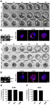
Figure 2. Homozygous NcEc embryos fail to form a functional trophectoderm.
(A) Still images from a twenty-four-hour time-lapse recording of in vitro cultured embryos from heterozygous EcNc intercrosses at E2.5. All embryos properly compact and separate the ICM (white arrow) from the TE (yellow arrowheads). (B) PCR genotyping of embryos in (A) and HA.11 immunofluorescence labeling of EcNc protein given in surface and medial optical sections, as well as z-stack 3D reconstructions, demonstrating the proper distribution of EcNc at the basolateral membrane (arrowheads). (C) Still images from a twenty-four-hour time-lapse recording of in vitro cultured embryos from heterozygous NcEc intercrosses at E2.5. Compaction is accomplished in all genotypes, but NcEc homozygous embryos cannot form a TE layer. (D) Similar analysis of NcEc homozygous embryos as shown in (B). Although NcEc is located mainly at basolateral sites (arrowheads), a uniform distribution was found in several outer cells (yellow arrow), and fragmented nuclei (white arrows) were also detected. (E,F) Percentage of embryos that formed a proper blastocyst in time-lapse experiments and during in vitro culture for EcNc (E, n = 33 wt, n = 101 ki/+ and n = 48 ki/ki) and NcEc mutants (F, n = 58 wt, n = 156 ki/+ and n = 56 ki/ki) in >10 independent experiments. Scale bar, 25 µm.
Figure 3
Figure 3. Markers for lineage specification, cell polarity, and vectorial fluid flow are correctly expressed and localized in NcEc homozygous mutants.
(A) Proper segregation of outer TE cells is shown by anti-Cdx2 labeling in wt, EcNc and NcEc homozygous mutants at E3.5. (B) Wildtype, EcNc and NcEc homozygous mutant embryos labeled for the ICM cell marker Sox2 in inner cells, showing ICM cell specification and its localization inside NcEc homozygous embryos. (C) Ezrin and Na+/K+-ATPase staining to verify apical-basal polarity with same distribution in wt, EcNc and NcEc homozygous mutants at the apical and basolateral membrane. Correct sealing of the TE layer is indicated by the presence and proper localization of the tight junctional component ZO-1 at apical sites of lateral TE membranes in wt, EcNc and NcEc homozygous embryos. Key components required for vectorial fluid transport are shown by the presence of Na+/K+-ATPase and Aqp3. In embryos of all genotypes, this expression is detected in the outer cells, without any obvious differences in expression between the embryos. Hence, the first lineage segregation is specified correctly, and proteins that are essential for the TE formation process and its function are present and properly localized in NcEc homozygous mutants. Nuclei were labeled with DAPI (blue). Scale bar, 25 µm.
Figure 4
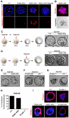
Figure 4. Increased apoptosis is detected in the outer cells of homozygous NcEc embryos and is blocked by iloprost treatment, rescuing blastocyst formation.
(A) Labeling of cleaved and activated Caspase 3 (red) shows only one apoptotic cell in the ICM of control and EcNc ki/ki embryos, whereas the outer cells of NcEc homozygous mutant embryos display a substantial increase in Caspase 3-positive cells. Cleavage of Caspase 3 was not detected upon iloprost treatment (1 µM) (B) Cell blebbing and vacuolization is detected in TROMA-1 positive outer cells of NcEc ki/ki embryos demonstrating induction of PCD in cells destined to become TE (arrow). (C) Treatment of NcEc ki/ki embryos with 1 µM iloprost at the precompacted or compacted morula stage (E2.25–E3.0) observed by time-lapse microscopy rescues the TE formation defect and hatching is initiated (arrow). (D) Treatment with iloprost at a later time-point (E3.5) did not rescue the phenotype. (E) One representative frame of a time-lapse recording of iloprost-treated EcNc ki/ki embryos. The formation of the blastocyst is marginally improved. (F) Treatment of N-cad ki/ki embryos with iloprost resulted in a rescue similar to that for NcEc homozygous mutants. (G) E-cad-null embryos were not rescued upon treatment with iloprost. (H) Percentage of iloprost-treated embryos that formed a proper blastocyst in time-lapse experiments and during in vitro culture for wt (n = 65), NcEc homozygous mutants (n = 33) and E-cad−/− (n = 15) in >5 independent experiments. (I) With the exception of E-cad−/− embryos, proper specification of the rescued TE of iloprost-treated homozygous mutants was confirmed by cytokeratin 8 expression, which is restricted to TE cells (TROMA-1, red). Scale bar, 25 µm.
Figure 5
Figure 5. Artificial increase of Igf1 levels during in vitro culture rescues blastocyst formation suggesting Igf1 signaling as the endogenous prosurvival stimulus.
(A) Wildtype and homozygous NcEc mutant embryos were incubated in the presence of 100 ng/ml Igf1 and recorded in 15-min intervals for 24 h. Images are displayed for 6-h intervals. Mutant embryos form a proper blastocyst similar to their control littermates and initiation of hatching is observed (arrow). (B) Insulin treatment rescues TE formation in a similar but milder fashion compared to Igf1 treatment. (C) Incubation of homozygous EcNc mutant embryos with Igf1 did not result in significant changes in blastocyst formation. (D) N-cad ki/ki embryos formed a blastocyst in the presence of Igf1. (E) E-cad−/− embryos were not rescued by Igf1 treatment. (F) Percentage of Igf1-treated embryos that formed a proper blastocyst in time-lapse experiments and during in vitro culture for wt (n = 31), NcEc homozygous mutants (n = 18) and E-cad−/− (n = 13) in >5 independent experiments. (G) Incubation of wt embryos with 100 ng/ml Igf1 or 10 µm Tyrphostin AG1024, a specific Igf1r inhibitor, induced hyperactivation or absence of Igf1r activation, respectively, demonstrating specificity of both the anti-Igf1r antibody and the inhibitor. Nuclear staining of the β-subunit and the phosphorylated form of Igf1r is additionally detected in the nucleus and is increasing upon Igf1 treatment as observed previously . (H) The activated form of Igf1r showed protein colocalization with E-cad in the TE cells of wt embryos. An antibody detecting the α- or the β-subunit of Igf1r (total Igf1r) showed localization of Igf1r throughout the membrane, partially overlapping with E-cad (left and middle panel, respectively). A complete overlap of the activated phosphorylated form of the receptor (pIgf1r) and E-cad labeling at lateral cell-cell contact sites was detected in the TE (right panel). (I) Igf1r was hypoactivated in NcEc- and E-cad-null embryos. Immunofluorescence labeling of pIgf1r and total Igf1r showed comparable intensities of activated Igf1r in wt and EcNc embryos, whereas a substantial reduction was found in NcEc and E-cad−/− embryos. Total Igf1r levels were unaffected. Scale bar, 25 µm.
Figure 6
Figure 6. E-cad interacts with Igf1r and increases receptor activity in TE cells.
(A) A proximity ligation assay (Duolink) examining wt (1, 4, 6), E-cad-null (2, 5) and NcEc homozygous embryos (3). Red dotted fluorescence indicates sites of interaction of analyzed proteins as indicated in optical sections (lower row) and 3D reconstructions (upper row). The E-cad-Igf1r interaction was detected by antibodies against the β-subunit of Igf1r and the intracellular domain of E-cad, which also binds to the NcEc protein. In wt embryos, fluorescent dots indicating sites of protein-protein interactions, were found at cell-cell contact sites (1), whereas E-cad-null embryos that contained only residual maternally derived E-cad and NcEc homozygous embryos did not show a fluorescence signal, although both anti-Igf1r and anti-E-cad antibodies detect the β-subunit of Igf1r and NcEc, respectively in NcEc homozygous mutant embryos (2, 3). Similar results were obtained in a Duolink assay using anti-E-cad (intracellular domain) and anti-pIgf1r (activated form) antibodies (4, 5). The known interaction between E-cad and β-catenin gave a punctuate pattern at basolateral membranes in cells of wt embryos as control (6). (B) The interaction between E-cad and Igf1r analyzed by co-immunoprecipitation experiments in wt and NcEc ki/ki TS cells. Immunoprecipitation with anti-E-cad and IgG control antibodies displayed a specific interaction of E-cad to coprecipitated Igf1r (total) in wt TS cells (upper panel) whereas the receptor was not co-immunoprecipitated with the NcEc protein in NcEc ki/ki TS cells (lower panel). 5% input was loaded in the last lane. Scale bar, 25 µm.
Figure 7
Figure 7. A model of the molecular pathways that are involved in TE survival but are blocked in homozygous NcEc and N-cad ki/ki mutants.
In the presence of full-length E-cad or of the E-cad extracellular domain in EcNc embryos interaction of cadherins with Igf1r is occurring. This enables proper activation of Igf1r upon Igf1 signaling (phosphorylation), which supplies survival signals and blocks PCD (left panel). In the absence of E-cad cell adhesion is maintained in presence of NcEc or N-cad, but both proteins are incapable of interacting with Igf1r. As a consequence of the uncoupled interaction, Igf1r is not fully activated, prosurvival signals are lacking and apoptotic pathways reach the threshold levels for PCD induction (right panel). In the presence of cadherin-mediated adhesion (in homozygous NcEc and N-cad ki/ki, but not in E-cad-null embryos), apoptotic pathways can be blocked only by external cues (red boxes), which inhibit PCD at different levels and thereby rescue TE formation. According to this model E-cad is required for providing survival cues via the extracellular domain in addition to its role in cell adhesion.

References

    1. Cockburn K, Rossant J. Making the blastocyst: lessons from the mouse. J Clin Invest. 2010;120:995–1003. - PMC - PubMed
    1. Rossant J, Tam PP. Blastocyst lineage formation, early embryonic asymmetries and axis patterning in the mouse. Development. 2009;136:701–713. - PubMed
    1. Avilion AA, Nicolis SK, Pevny LH, Perez L, Vivian N, et al. Multipotent cell lineages in early mouse development depend on SOX2 function. Genes Dev. 2003;17:126–140. - PMC - PubMed
    1. Chazaud C, Yamanaka Y, Pawson T, Rossant J. Early lineage segregation between epiblast and primitive endoderm in mouse blastocysts through the Grb2-MAPK pathway. Dev Cell. 2006;10:615–624. - PubMed
    1. Nichols J, Zevnik B, Anastassiadis K, Niwa H, Klewe-Nebenius D, et al. Formation of pluripotent stem cells in the mammalian embryo depends on the POU transcription factor Oct4. Cell. 1998;95:379–391. - PubMed

Publication types

MeSH terms