. 2012 Mar 29:3:116.
doi: 10.3389/fmicb.2012.00116. eCollection 2012.

Carbohydrate Derived Fulvic Acid: An in vitro Investigation of a Novel Membrane Active Antiseptic Agent Against Candida albicans Biofilms

Affiliations

Carbohydrate Derived Fulvic Acid: An in vitro Investigation of a Novel Membrane Active Antiseptic Agent Against Candida albicans Biofilms

Leighann Sherry et al. Front Microbiol. .

Abstract

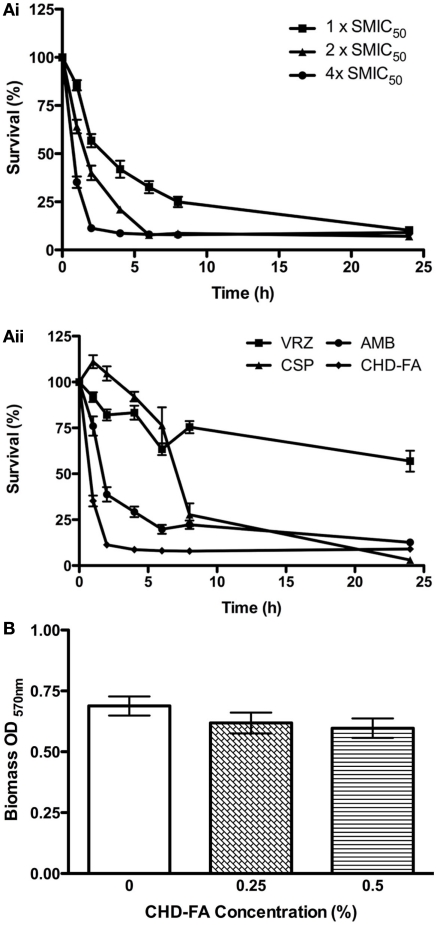
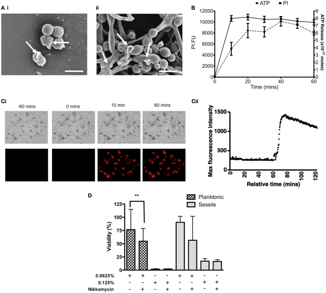
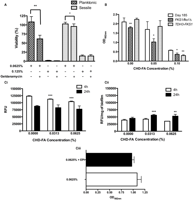
Carbohydrate derived fulvic acid (CHD-FA) is a heat stable low molecular weight, water soluble, cationic, colloidal material with proposed therapeutic properties. The aim of this study was to evaluate the antifungal activity of CHD-FA against Candida albicans, and to characterize its mode of action. A panel of C. albicans isolates (n = 50) derived from a range of clinical specimens were grown planktonically and as biofilms, and the minimum inhibitory concentrations determined. Scanning electron microscopy was performed to examine ultrastructural changes and different cell membrane assays were used to determine its mode of action. In addition, the role of C. albicans biofilm resistance mechanisms were investigated to determine their effects on CHD-FA activity. CHD-FA was active against planktonic and sessile C. albicans at concentrations 0.125 and 0.25% respectively, and was shown to be fungicidal, acting through disruption of the cell membrane activity. Resistance mechanisms, including matrix, efflux, and stress, had a limited role upon CHD-FA activity. Overall, based on the promising in vitro spectrum of activity and minimal biofilm resistance of the natural and cheap antiseptic CHD-FA, further studies are required to determine its applicability for clinical use.

Keywords: Candida albicans; antiseptic; biofilm; fulvic acid.


Figures

Figure 1
Figure 1
Candida albicans biofilm killing by CHD-FA is time and concentration dependent, despite no biofilm disruption. (A) Biofilm time-kill kinetics of CHD-FA at 1, 2, and 4 × SMIC50. (i) Biofilm time-kill kinetics of VRZ, CSP, AMB and CHD-FA at 4 × SMIC50. (ii) Standardized C. albicans (1 × 106 cells/ml) were grown in flat-bottomed 96-well plates for 24 h, washed in PBS and treated with antifungal agents at defined concentrations for 1, 2, 4, 6, 8, and 24 h. Metabolic activity of treated biofilms was then quantified using the XTT assay. Four isolates were used for each assay, and this was performed on two independent occasions in triplicate. (B) Standardized C. albicans (1 × 106 cells/ml) were grown in flat bottomed 96-well plates for 24 h, washed in PBS and treated with 0.25 and 0.5% CHD-FA for 24 h. A negative control was also included. The biomass of the biofilm was then quantified by staining each biofilm with 0.05% w/v crystal violet solution. The biofilms were washed, allowed to air dry, and 100% ethanol applied to destain each biofilm. The biomass was quantified spectrophotometrically by reading at 570 nm in a microtiter plate reader (FLUOStar Omega, BMG Labtech). Forty isolates were tested during this assay, which was performed in triplicate.
Figure 2
Figure 2
Carbohydrate derived fulvic acid permeabilizes Candida albicans cell membranes. (A) Planktonic cells (i) and sessile (ii) cells were treated at their respective MIC90 for 24 h either in solution or on Thermanox™ coverslips, respectively. These were then processed and viewed on a JEOL JSM-6400 scanning electron microscope and images assembled using Photoshop software. Note the apparent disruption of the cell wall denoted by arrows on both the planktonic and sessile cells. Scale bars represent 5 μm. (B) C. albicans (SC5314) planktonic cells (5 × 107 cells/ml) were treated with CHD-FA (4%) for 10, 20, 30, 40, 50, and 60 min. A negative control (no CHD-FA) was also included. For PI experiments the cells were washed by centrifugation, resuspended in PI (20 μM in PBS), and incubated for 15 min at 37°C. These were then transferred to a black 96-well plate for quantification in a fluorescent plate reader (Ex485/Em620). For ATP release the cells were removed by filter sterilization (0.22 μm) and supernatants adjusted to a pH of 7.8 in 10 mM NaOH. The assay, including standards, was performed as per manufacturer’s instructions in a white well plate for quantification in a luminescent plate reader. Each assay was performed on at least two independent occasions in triplicate. (C) C. albicans cells were allowed to grow on an Ibidi μSlide VI for 60 min before 20 μM PI and 2% CHD-FA was added. PI uptake was measured over 60 min using time-lapse microscopy, with images taken every 20 s. Note the sudden uptake of PI into C. albicans cells after exposure to CHD-FA for 10 min (i), which was validated through quantifiable data that showed a rapid increase in fluorescence when CHD-FA was added (ii). (D) C. albicans was grown as planktonic and biofilm cells ± nikkomycin Z (0.4 μg/ml) for 24 h. These were then treated with 0.0625 and 0.125% CHD-FA for 24 h. Negative controls were included. Metabolic activity of treated cells was then quantified using the XTT assay. Four isolates were used for each assay, and this was performed on two independent occasions in quadruplicate. *p < 0.05, **p < 0.01, ***p < 0.001.
Figure 3
Figure 3
Candida albicans biofilm resistance mechanisms play a limited role in CHD-FA sensitivity. (A) C. albicans was grown as planktonic and biofilm cells ± geldanamycin (12.5 μg/ml) for 24 h. These were then treated with 0.0625 and 0.125% CHD-FA for 24 h. Negative controls were included. Metabolic activity of treated cells was then quantified using the XTT assay. Four isolates were used for each assay, and this was performed on two independent occasions in quadruplicate. (B) C. albicans DAY185, FKS1/fks1Δ and TDH3-FKS1 were grown as biofilms for 8 h and treated with 0.05 and 0.1% CHD-FA for 24 h. Negative controls were included. Metabolic activity of treated cells was then quantified using the XTT assay. This was performed on two independent occasions in triplicate. Statistical significance represents the comparison of FKS1/fks1Δ to the reference strain DAY185. (C) C. albicans SC5314 was grown as (i) planktonic and (ii) biofilm cells and treated with 0.0313 and 0.0625% CHD-FA (sub-MIC levels) for 4 and 24 h. Cells were washed, resuspended in assay buffer and 100 μg/ml of Ala-Nap added. These were then transferred to a black 96-well plate for quantification at 30 s intervals over 60 min in a fluorescent plate reader (Ex355/Em460). For biofilms the relative fluorescence is presented, which is normalized to dry weight of biofilms (RFU/mg). High fluorescence values indicate low efflux activity and vice versa. C. albicans 24 h biofilms were treated with 0.0625% CHD-FA ± EPI for 24 h (iii). Biofilms were washed and metabolic activity measured using the XTT metabolic assay with absorbance read at 492 nm. *p < 0.05, **p < 0.01, ***p < 0.001.

References

    1. Ahmad A., Khan A., Kumar P., Bhatt R. P., Manzoor N. (2011). Antifungal activity of Coriaria nepalensis essential oil by disrupting ergosterol biosynthesis and membrane integrity against Candida. Yeast 28, 611–617 10.1002/yea.1890 - DOI - PubMed
    1. Baillie G. S., Douglas L. J. (2000). Matrix polymers of Candida biofilms and their possible role in biofilm resistance to antifungal agents. J. Antimicrob. Chemother. 46, 397–403 10.1093/jac/46.3.397 - DOI - PubMed
    1. Clinical Laboratory Standards Institute (CLSI) (2008). Reference Method for Broth Dilution Antifungal Susceptibility Testing of Yeast, Approved Standard – 3rd Edn, CLSI document M27-A3. Wayne, PA: CLSI
    1. Coco B. J., Bagg J., Cross L. J., Jose A., Cross J. G., Ramage G. (2008). Mixed Candida albicans and Candida glabrata populations associated with the pathogenesis of denture stomatitis. Oral Microbiol. Immunol. 23, 377–383 10.1111/j.1399-302X.2008.00439.x - DOI - PubMed
    1. Dusane D. H., Rajput J. K., Kumar A. R., Nancharaiah Y. V., Venugopalan V. P., Zinjarde S. S. (2008). Disruption of fungal and bacterial biofilms by lauroyl glucose. Lett. Appl. Microbiol. 47, 374–379 10.1111/j.1472-765X.2008.02440.x - DOI - PubMed

LinkOut - more resources