. 2012;7(3):e33066.
doi: 10.1371/journal.pone.0033066. Epub 2012 Mar 30.

Composition and evolution of the vertebrate and mammalian selenoproteomes

Affiliations

Composition and evolution of the vertebrate and mammalian selenoproteomes

Marco Mariotti et al. PLoS One. 2012.

Abstract

Background: Selenium is an essential trace element in mammals due to its presence in proteins in the form of selenocysteine (Sec). Human genome codes for 25 Sec-containing protein genes, and mouse and rat genomes for 24.

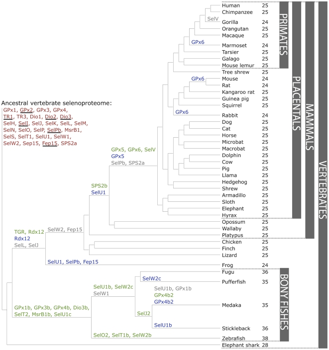
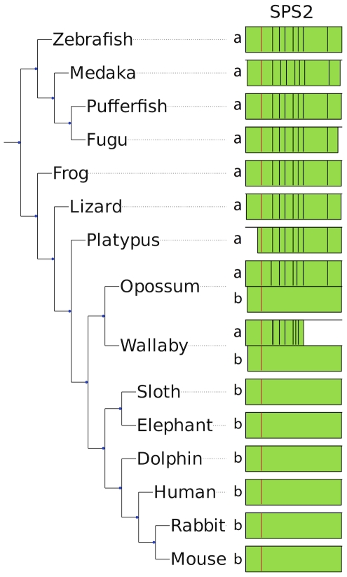
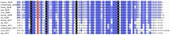
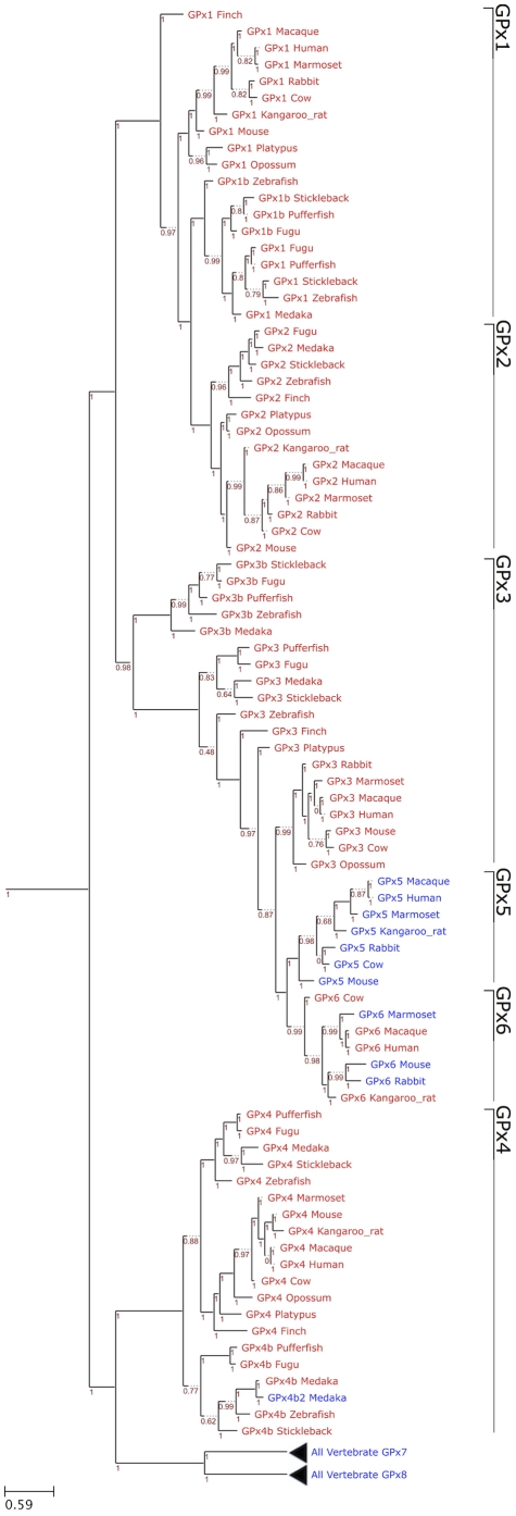
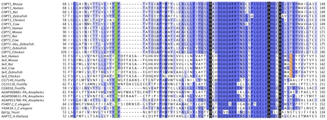
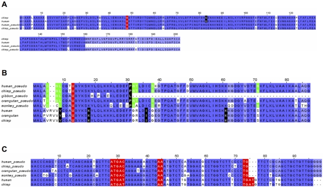
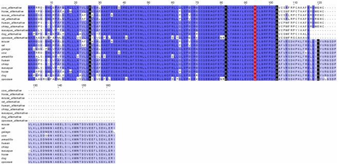
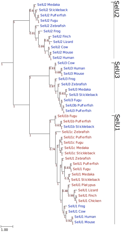
Methodology/principal findings: We characterized the selenoproteomes of 44 sequenced vertebrates by applying gene prediction and phylogenetic reconstruction methods, supplemented with the analyses of gene structures, alternative splicing isoforms, untranslated regions, SECIS elements, and pseudogenes. In total, we detected 45 selenoprotein subfamilies. 28 of them were found in mammals, and 41 in bony fishes. We define the ancestral vertebrate (28 proteins) and mammalian (25 proteins) selenoproteomes, and describe how they evolved along lineages through gene duplication (20 events), gene loss (10 events) and replacement of Sec with cysteine (12 events). We show that an intronless selenophosphate synthetase 2 gene evolved in early mammals and replaced functionally the original multiexon gene in placental mammals, whereas both genes remain in marsupials. Mammalian thioredoxin reductase 1 and thioredoxin-glutathione reductase evolved from an ancestral glutaredoxin-domain containing enzyme, still present in fish. Selenoprotein V and GPx6 evolved specifically in placental mammals from duplications of SelW and GPx3, respectively, and GPx6 lost Sec several times independently. Bony fishes were characterized by duplications of several selenoprotein families (GPx1, GPx3, GPx4, Dio3, MsrB1, SelJ, SelO, SelT, SelU1, and SelW2). Finally, we report identification of new isoforms for several selenoproteins and describe unusually conserved selenoprotein pseudogenes.

Conclusions/significance: This analysis represents the first comprehensive survey of the vertebrate and mammal selenoproteomes, and depicts their evolution along lineages. It also provides a wealth of information on these selenoproteins and their forms.


Conflict of interest statement

Competing Interests: The authors have declared that no competing interests exist.

Figures

Figure 1
Figure 1. Evolution of the vertebrate selenoproteome.
The ancestral vertebrate selenoproteome is indicated in red, and its changes across the investigated vertebrates are depicted along their phylogenetic tree. The ancestral selenoproteins found uniquely in vertebrates are underlined. The creation of a new selenoprotein (here always by duplication of an existing one) is indicated by its name in green. Loss is indicated in grey. Replacement of Sec with Cys is indicated in blue (apart from SelW2c in pufferfish, which is with arginine). Events of conversion of Cys to Sec were not found. On the right, the number of selenoproteins predicted in each species is shown.
Figure 2
Figure 2. Replacement of a multiexon SPS2a by an intronless SPS2b.
In the figure, the SPS2 genes found in some representative species are shown. The positions of introns along the protein sequence are displayed with black lines, and the Sec residue is displayed in red. In a few cases, the predicted genes were incomplete because of poor sequence data (e.g., the N-terminal region in platypus). Placental mammals (bottom) possess a single intronless gene, SPS2b. Non-mammalian vertebrates (top) and platypus possess a single multiexon gene, SPS2a. Marsupials (opossum and wallaby) possess both.
Figure 3
Figure 3. Multiple sequence alignment of SelV and SelW.
The last 9 residues of SelV exon 1 and exons 2–5 are shown aligned to complete SelW sequences. The last residue of each exon is marked in black and the Sec in red.
Figure 4
Figure 4. Phylogenetic tree of GPx family in eukaryotes.
The figure shows a ML tree computed using the JTT substitution model. In the phylogram, Sec-containing proteins are shown in red and Cys-containing homologs are shown in blue. The GPx families are indicated on the right. The distance scale in substitutions per position is indicated at the bottom left. The branch support is shown in red.
Figure 5
Figure 5. Multiple sequence alignment of SelI and its homologs.
The multiple sequence alignment of the active site and preceding regions of CHPT1, CEPT1, and SelI is shown. Homologs are labeled with the annotated name. Proteins in the bottom section comprise a large group of diverse proteins containing the same domain. The most critical residues are marked in red. The residue in green marks the end of the first transmembrane domain. The cysteine residue near the active site emerged specifically in SelI proteins is marked in orange.The full length alignment is provided in Supplementary Figure S37 and the corresponding phylogenetic tree in Supplementary Figure S38.
Figure 6
Figure 6. SECIS elements of SelM and SelO.
Multiple sequence alignment of SelM (A) and SelO (B) SECIS elements. Critical regions are marked in red.
Figure 7
Figure 7. Multiple sequence alignment of selenoprotein genes and pseudogenes.
A. GPx1. Multiple sequence alignment of human and chimpanzee GPx1 pseudogenes. B. SelW. The last residue of each exon is marked in black and Sec in red. Residues marked in green are described in the text. C. SECIS elements of SelW and SelW pseudogene.
Figure 8
Figure 8. Multiple sequence alignment of Sep15 and a Sep15 alternative isoform.
The last residue of each exon is marked in black and Sec in red. For human and mouse, ESTs support the presence of the isoform. For the other species shown, the protein sequences were predicted simulating skipping the 4th exon.
Figure 9
Figure 9. Phylogeny of SelU family in vertebrates.
ML tree computed using the JTT substitution model. Sec-containing proteins are shown in red, whereas the Cys-containing homologs are shown in blue. At the bottom left, the distance scale in substitutions per position is shown. Branch support is shown along the tree in red.

References

    1. Böck A, Forchhammer K, Heider J, Leinfelder W, Sawers G, et al. Selenocysteine: the 21st amino acid. Mol Microbiol. 1991;5:515–520. - PubMed
    1. Stadtman TC. Selenocysteine. Annu Rev Biochem. 1996;65:83–100. - PubMed
    1. Hatfield DL, Gladyshev VN. How selenium has altered our understanding of the genetic code. Mol Cell Biol. 2002;22:3565–3576. - PMC - PubMed
    1. Berry MJ, Banu L, Chen YY, Mandel SJ, Kieffer JD, et al. Recognition of UGA as a selenocysteine codon in type I deiodinase requires sequences in the 3′ untranslated region. Nature. 1991;353:273–276. - PubMed
    1. Low SC, Berry MJ. Knowing when to stop: selenocysteine incorporation in eukaryotes. Trends Biochem Sci. 1996;21:203–208. - PubMed

Publication types

MeSH terms