. 2012;7(3):e33163.
doi: 10.1371/journal.pone.0033163. Epub 2012 Mar 30.

Granzyme B cleaves decorin, biglycan and soluble betaglycan, releasing active transforming growth factor-β1

Affiliations

Granzyme B cleaves decorin, biglycan and soluble betaglycan, releasing active transforming growth factor-β1

Wendy A Boivin et al. PLoS One. 2012.

Erratum in

  • PLoS One. 2012;7(5): doi/10.1371/annotation/b1e4ff60-ba18-4f92-b856-0f2dd27e9a65

Abstract

Objective: Granzyme B (GrB) is a pro-apoptotic serine protease that contributes to immune-mediated target cell apoptosis. However, during inflammation, GrB accumulates in the extracellular space, retains its activity, and is capable of cleaving extracellular matrix (ECM) proteins. Recent studies have implicated a pathogenic extracellular role for GrB in cardiovascular disease, yet the pathophysiological consequences of extracellular GrB activity remain largely unknown. The objective of this study was to identify proteoglycan (PG) substrates of GrB and examine the ability of GrB to release PG-sequestered TGF-β1 into the extracellular milieu.

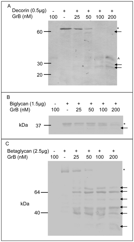
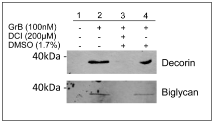
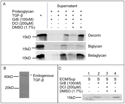
Methods/results: Three extracellular GrB PG substrates were identified; decorin, biglycan and betaglycan. As all of these PGs sequester active TGF-β1, cytokine release assays were conducted to establish if GrB-mediated PG cleavage induced TGF-β1 release. Our data confirmed that GrB liberated TGF-β1 from all three substrates as well as from endogenous ECM and this process was inhibited by the GrB inhibitor 3,4-dichloroisocoumarin. The released TGF-β1 retained its activity as indicated by the induction of SMAD-3 phosphorylation in human coronary artery smooth muscle cells.

Conclusion: In addition to contributing to ECM degradation and the loss of tissue structural integrity in vivo, increased extracellular GrB activity is also capable of inducing the release of active TGF-β1 from PGs.


Conflict of interest statement

Competing Interests: The authors have read the journal's policy and have the following conflicts: DJG is a Founder and Scientific Officer to viDA Therapeutics, Inc., however, viDA was not involved in any aspect of this study. No commercial/therapeutic products from viDA were used in this study. This does not alter the authors' adherence to all the PLoS ONE policies on sharing data and materials.

Figures

Figure 1
Figure 1. GrB-mediated cleavage of decorin, biglycan and betaglycan.
Increasing concentrations of GrB (25, 50, 100 and 200 nM) were incubated with decorin (a), biglycan (b), and betaglycan (c) for 24 h at RT. * denotes full length protein, arrows indicate cleavage fragments and ∧ indicates GrB.
Figure 2
Figure 2. GrB-mediated PG cleavage is inhibited by DCI and cleavage site identification.
GrB was incubated with decorin (a), biglycan (b) and betaglycan (c), +/− DCI and the solvent control DMSO, for 4 h and 24 h. Cleavage sites in biglycan and betaglycan were identified by N-terminal Edman degradation. * denotes full length protein, arrows indicate cleavage fragments, and cleavage sites are displayed on the right.
Figure 3
Figure 3. GrB cleaves native smooth muscle cell-derived decorin and biglycan.
HCASMCs were incubated at confluency for adequate ECM synthesis. Cells were removed, GrB was incubated with the ECM and decorin and biglycan cleavage fragments were detected by western immunoblotting.
Figure 4
Figure 4. GrB-mediated cleavage of decorin, biglycan and betaglycan results in the release of active TGF-β1.
Decorin, biglycan and betaglycan complexed with TGF-β1 were treated with GrB. Supernatants (containing released TGF-β1), were collected and released TGF-β1 was detected by Western blotting. Results shown are representative western blots from at least 3 separate experiments for each PG (a). As endogenous SMC-derived ECM only contains latent TGF-β (as shown in (b)), GrB-mediated release from active TGF-β1 supplemented ECM was also examined (c).
Figure 5
Figure 5. TGF-β1 released by GrB is active and induces SMAD-3 activation in HCASMCs.
GrB+/−DCI was incubated on betaglycan/TGF-β1 complexes for 24 h. Supernatants (containing released TGF-β1) were added to HCASMC for 20 m and phosphorylated SMAD-2 and SMAD-3 levels were examined. TGF-β1 released by GrB is active and induces SMAD-3 signalling in HCASMCs (P<0.05). The result shown is representative of at least 5 experiments.

References

    1. Medema JP, Toes RE, Scaffidi C, Zheng TS, Flavell RA, et al. Cleavage of FLICE (caspase-8) by granzyme B during cytotoxic T lymphocyte-induced apoptosis. Eur J Immunol. 1997;27:3492–3498. - PubMed
    1. Boivin WA, Cooper DM, Hiebert PR, Granville DJ. Intracellular versus extracellular granzyme B in immunity and disease: challenging the dogma. Lab Invest. 2009;89:1195–1220. - PMC - PubMed
    1. Hendel A, Hiebert PR, Boivin WA, Williams SJ, Granville DJ. Granzymes in age-related cardiovascular and pulmonary diseases. Cell Death Differ. 2010;17:596–606. - PubMed
    1. Bratke K, Bottcher B, Leeder K, Schmidt S, Kupper M, et al. Increase in granzyme B+ lymphocytes and soluble granzyme B in bronchoalveolar lavage of allergen challenged patients with atopic asthma. Clin Exp Immunol. 2004;136:542–548. - PMC - PubMed
    1. Spaeny-Dekking EH, Hanna WL, Wolbink AM, Wever PC, Kummer AJ, et al. Extracellular granzymes A and B in humans: detection of native species during CTL responses in vitro and in vivo. J Immunol. 1998;160:3610–3616. - PubMed

Publication types

MeSH terms