. 2012;7(3):e33876.
doi: 10.1371/journal.pone.0033876. Epub 2012 Mar 27.

High resolution intravital imaging of subcellular structures of mouse abdominal organs using a microstage device

Affiliations

High resolution intravital imaging of subcellular structures of mouse abdominal organs using a microstage device

Liqin Cao et al. PLoS One. 2012.

Abstract

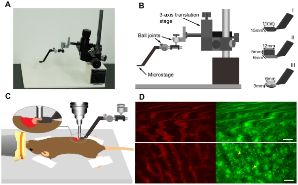
Intravital imaging of brain and bone marrow cells in the skull with subcellular resolution has revolutionized neurobiology, immunology and hematology. However, the application of this powerful technology in studies of abdominal organs has long been impeded by organ motion caused by breathing and heartbeat. Here we describe for the first time a simple device designated 'microstage' that effectively reduces organ motions without causing tissue lesions. Combining this microstage device with an upright intravital laser scanning microscope equipped with a unique stick-type objective lens, the system enables subcellular-level imaging of abdominal organs in live mice. We demonstrate that this technique allows for the quantitative analysis of subcellular structures and gene expressions in cells, the tracking of intracellular processes in real-time as well as three-dimensional image construction in the pancreas and liver of the live mouse. As the aforementioned analyses based on subcellular imaging could be extended to other intraperitoneal organs, the technique should offer great potential for investigation of physiological and disease-specific events of abdominal organs. The microstage approach adds an exciting new technique to the in vivo imaging toolbox.


Conflict of interest statement

Competing Interests: The authors have read the journal's policy and have the following conflicts: L.C. and K.A. filed a patent application covering the microstage device (No. 2008-100505). This does not alter the authors' adherence to all the PLoS ONE policies on sharing data and materials.

Figures

Figure 1
Figure 1. Effect of the microstage device on image stability and quality.
(A) Microstage device. (B) Illustration of the microstage device and microstages of three types. (C) Illustration showing that a portion of the pancreas of an anesthetized mouse was positioned on the microstage and imaged using a stick-type water immersion lens. (D) In vivo images of liver vasculature (left, red, labeled with AngioSense 750 IVM) and liver cells (right, green) of the GFP–LC3 mouse taken without (top row) and with (bottom row) the microstage device. Arrows depict GFP-labeled autophagic membranes. Individual images in D are 512×256 pixel areas cropped from 512×512 pixels. Scale bars, 20 µm.
Figure 2
Figure 2. In vivo imaging of subcellular structures and gene expression in mouse abdominal organs with the microstage device.
(A–D) Highly methylated chromocenters labeled by mRFP–MBD-nls in cells of liver (A), kidney (B), pancreas (C) and skeletal muscle (D) of mRFP–MBD-nls transgenic mice. Green pseudocolor shows the vasculatures labeled with an intravenous injection of AngioSense 750 IVM. Insets: magnified pictures of the boxed area in corresponding images. (E) Quantitative analysis of chromocenter numbers in cell nuclei of four types of tissues. Counts of 24, 23, 23 and 32 cell nuclei were analyzed for the liver, kidney, pancreas and skeletal muscle, respectively. (F–I) Autophagy in pancreatic acinar cells (F, G) and skeletal muscle (H, J) of GFP–LC3 mice before (F, H) and after (G, I) 24 h starvation. (J) Quantitative analysis of autophagy in response to 24 h starvation. The numbers of GFP–LC3 dots were counted and divided by the corresponding area (n = 5 image fields). (K–R) Fluorescence intensity in liver (K, O), kidney (L, P), pancreas (M, Q) and skeletal muscle (N, R) of ERAI-transgenic mice injected with saline (K–N) or tunicamycin (O–R). (S) Quantitative analysis of fluorescence intensity in tissues under different physiological conditions (n = 3 image fields). Results in E, J and S represent the mean ± SD. **P<0.01. Images in A–D and F–I are 512×512 pixels. Images in K–R are 320×320 pixels. Scale bars, 20 µm (A–D, F–I) and 50 µm (K–R).
Figure 3
Figure 3. In vivo imaging of cellular processes in the pancreas of live GFP–LC3 mice starved for 24 h (see also Movie S5).
(A) Pancreatic acinar cells (left) and vasculature (right). (B) Time-lapse images of autophagosome movements in the boxed region (yellow) in A at the indicated times. Two GFP–LC3 dots exhibited distinct behaviors; one showed rapid displacement (yellow arrow), while the other remained stationary (yellow arrowhead). (C) Time-lapse images of an event similar to exocytosis in the boxed region (magenta) in A at the indicated times. Magenta arrows designate the protrusion-like profile. (D) Time-lapse images of pancreatic microcirculation obtained from the boxed region (white) in A at the indicated times. Individual cells appeared as dark foci in the vasculature. White arrowheads mark progress of a single cell. Scale bars, 20 µm (A), 2 µm (B) and 5 µm (C, D).

References

    1. Drew PJ, Shih AY, Driscoll JD, Knutsen PM, Blinder P, et al. Chronic optical access through a polished and reinforced thinned skull. Nat Methods. 2010;7:981–984. - PMC - PubMed
    1. Flusberg BA, Nimmerjahn A, Cocker ED, Mukamel EA, Barretto RP, et al. High-speed, miniaturized fluorescence microscopy in freely moving mice. Nat Methods. 2008;5:935–938. - PMC - PubMed
    1. Looney MR, Thornton EE, Sen D, Lamm WJ, Glenny RW, et al. Stabilized imaging of immune surveillance in the mouse lung. Nat Methods. 2011;8:91–96. - PMC - PubMed
    1. Weigert R, Sramkova M, Parente L, Amornphimoltham P, Masedunskas A. Intravital microscopy: a novel tool to study cell biology in living animals. Histochem Cell Biol. 2010;133:481–491. - PMC - PubMed
    1. Murari K, Zhang Y, Li S, Chen Y, Li MJ, et al. Compensation-free, all-fiber-optic, two-photon endomicroscopy at 1.55 mum. Opt Lett. 2011;36:1299–1301. - PMC - PubMed

Publication types