. 2012;7(3):e34020.
doi: 10.1371/journal.pone.0034020. Epub 2012 Mar 30.

Xylosyltransferase-I regulates glycosaminoglycan synthesis during the pathogenic process of human osteoarthritis

Affiliations

Xylosyltransferase-I regulates glycosaminoglycan synthesis during the pathogenic process of human osteoarthritis

Narayanan Venkatesan et al. PLoS One. 2012.

Abstract

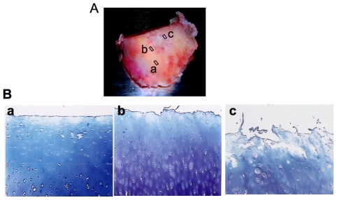
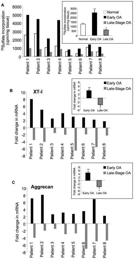
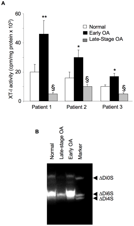
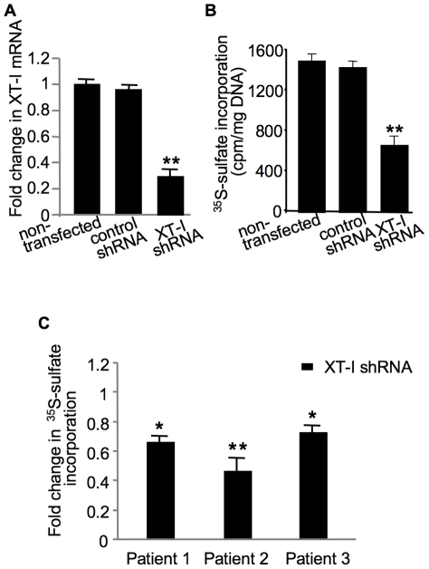
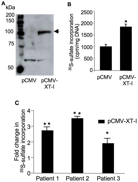
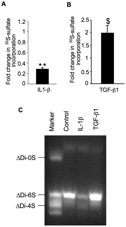
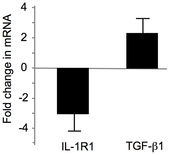
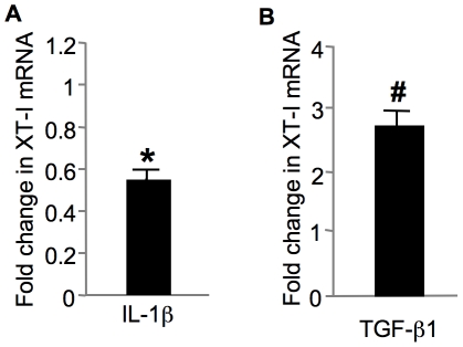
Loss of glycosaminoglycan (GAG) chains of proteoglycans (PGs) is an early event of osteoarthritis (OA) resulting in cartilage degradation that has been previously demonstrated in both huma and experimental OA models. However, the mechanism of GAG loss and the role of xylosyltransferase-I (XT-I) that initiates GAG biosynthesis onto PG molecules in the pathogenic process of human OA are unknown. In this study, we have characterized XT-I expression and activity together with GAG synthesis in human OA cartilage obtained from different regions of the same joint, defined as "normal", "late-stage" or adjacent to "late-stage". The results showed that GAG synthesis and content increased in cartilage from areas flanking OA lesions compared to cartilage from macroscopically "normal" unaffected regions, while decreased in "late-stage" OA cartilage lesions. This increase in anabolic state was associated with a marked upregulation of XT-I expression and activity in cartilage "next to lesion" while a decrease in the "late-stage" OA cartilage. Importantly, XT-I inhibition by shRNA or forced-expression with a pCMV-XT-I construct correlated with the modulation of GAG anabolism in human cartilage explants. The observation that XT-I gene expression was down-regulated by IL-1β and up-regulated by TGF-β1 indicates that these cytokines may play a role in regulating GAG content in human OA. Noteworthy, expression of IL-1β receptor (IL-1R1) was down-regulated whereas that of TGF-β1 was up-regulated in early OA cartilage. Theses observations may account for upregulation of XT-I and sustained GAG synthesis prior to the development of cartilage lesions during the pathogenic process of OA.


Conflict of interest statement

Competing Interests: The authors have declared that no competing interests exist.

Figures

Figure 1
Figure 1. Representative sample of femoral condyle from human OA knee and toluidine blue stained sections.
(A) Femoral condyle from human OA knee joint indicating three sites biopsied for further biochemical and molecular analysis; (a) cartilage sample taken from macroscopically unaffected area and distant from the lesion was termed “normal” (b) cartilage sample isolated from areas immediately close to lesion was termed “Next to lesion”; (c) cartilage specimen excised from lesion was termed “late-stage OA”; Cartilage samples were isolated using a 6 mm-biopsy punch. (B) Representative photomicrographs of cartilage sections showing areas from where samples were recovered for toluidine blue staining; (a) cartilage section from a normal area showing a relatively smooth articular surface; (b) section from areas close to lesion showing dense staining for PGs in mid zone of the cartilage where chondrocyte proliferation and activation occurs; (c) cartilage section from lesions showing degraded articular surface, loss of PG staining and chondrocyte cloning (original magnification ×40).
Figure 2
Figure 2. Analysis of GAG synthesis and expression of XT-I and aggrecan genes.
(A) GAG synthesis analysis in cartilage from “normal”, early OA and late-stage OA areas for each patient studied by 35S-sulfate incorporation. Insert shows the mean of GAG synthesis rate values corresponding to different areas of cartilage sample from all patients. Values are mean ± SEM of 3 experiments per parameter, per joint, per patient. (B) and (C) Analyses of the expression of XT-I and aggrecan genes in cartilage from normal, early OA and late-stage OA areas for each patient. The expression level of the genes was analyzed by qPCR. Measurements were normalized to the control “normal”. Inserts in (B) and (C) show the mean of XT-I and aggrecan gene expression level values corresponding to different areas of cartilage sample from all patients. Values are mean ± SD of 3 experiments per parameter, per joint, per patient. **significantly (P<0.01) higher than normal and late groups; §significantly (P<0.05) higher than normal and late groups; *significantly (P<0.05) lower than normal.
Figure 3
Figure 3. Analysis of xylosyltransferase activity and GAG chain composition.
(A) xylosyltransferase activity from normal, early OA and late-stage OA cartilage of three representative patients. Data are mean ± SD of 3 experiments per parameter, per joint, per patient. **Significantly (P<0.01) higher than all groups; *significantly (P<0.01) higher than normal and late groups; §significantly (P<0.05) lower than normal. (B) GAG chain composition in human cartilage explants as determined by FACE. Proteinase K-digested cartilage explants containing equal amounts (5 µg) of GAGs, as determined by uronic acid assay from each group, were digested with chondroitinase ABC and the disaccharides obtained were fluorotagged and derivatized. Disaccharides were run on a monocomposition gel and gels are representative images from 3 experiments per parameter, per joint, per patient. Markers are ΔDi0S, nonsulfated chondroitin-unsaturated disaccharide; ΔDi4S, chondroitin-4-sulfated unsaturated disaccharide; ΔDi6S, chondroitin-6-sulfated unsaturated disaccharide.
Figure 4
Figure 4. Analysis of the effect of XT-I knockdown on GAG synthesis in chondrocytes and cartilage explants.
Cells were transfected with XT-I shRNA, or control shRNA or left untreated and then examined for mRNA expression (A) and GAG synthesis (B). Total RNA was isolated from cells of each group at 72 h posttransfection and XT-I expression level was determined by qPCR analysis. XT-I mRNA levels are represented as a ratio relative to those in negative controls or untreated controls. GAG synthesis was studied by 35S-sulfate incorporation. (C) Cartilage explants from normal region of human femoral condyle of three patients were transfected with XT-I shRNA or control shRNA and then GAG synthesis was analyzed by 35S-sulfate incorporation. Measurements were normalized to control shRNA. Data represent the mean ± SD of 3 independent observations for each group. **,*Significantly (P<0.01, P<0.05, respectively) lower than control shRNA.
Figure 5
Figure 5. Effect of XT-I overexpression on GAG anabolism in chondrocytes and cartilage.
(A) Immunoblot of XT-I protein in pCMV- or pCMV-XT-I-transfected chondrocytes 48 h after transfection. XT-I recombinant protein was indicated by the arrow. (B) GAG synthesis in primary human chondrocytes or (C) in cartilage explants from late OA region of human femoral condyle transfected with pCMV- (control) or pCMV-XT-I vector. XT-I- or mock-transfected cells or cartilage were labeled with 35S-sulfate, and the amount of 35S-labeled GAGs was measured 48 h posttransfection. Data are mean ± SD of 3 experiments per parameter, per joint, per patient. **,*Significantly (P<0.01, P<0.05, respectively) higher than controls.
Figure 6
Figure 6. Analysis of the effect of IL-1β and TGF-β1 on GAG synthesis and GAG chain composition.
Cartilage explants from normal regions of human femoral condyle were exposed to IL-1β (A) or TGF-β1 (B) for 24 h and then GAG synthesis was measured by 35S-sulfate incorporation. Measurements were normalized to control (non treated). Data are mean ± SD of 3 experiments per parameter, per joint, per patient. **Significantly (P<0.0018) lower than controls; $significantly (P<0.05) higher than controls. (C) Effect of IL-1β and TGF-β1 on GAG chain composition in human cartilage. Cartilage samples derived from normal regions of human femoral condyle were treated with cytokines for 12 h and analyzed by FACE. Gels are representative images from 3 experiments per parameter, per joint, per patient. Markers are ΔDi0S, nonsulfated chondroitin-unsaturated disaccharide; ΔDi4S, chondroitin-4-sulfated unsaturated disaccharide; ΔDi6S, chondroitin-6-sulfated unsaturated disaccharide.
Figure 7
Figure 7. Expression of pro-inflammatory and anti-inflammatory cytokines in human OA cartilage.
Total RNA was isolated from human cartilage samples obtained from early OA and late OA regions and the expression of target genes were analyzed by qPCR. Target genes in early OA were compared to late OA and expressed as fold induction. Values are mean ± SD of 3 experiments per parameter per joint per patient.
Figure 8
Figure 8. Effect of IL-1β (A) and TGF-β1 (B) on XT-I gene expression in normal articular cartilage.
Cartilage samples isolated from normal regions of human femoral condyle was exposed to IL-1β or TGF-β1 and then XT-I gene expression was analyzed by qPCR. Measurements were normalized to control (non treated). Each value represents the mean ± SD of 3 experiments per parameter per joint per patient. *Significantly (P<0.05) lower than control group; #significantly (P<0.01) higher than control group.

References

    1. Heinegard D, Saxne T. The role of the cartilage matrix in osteoarthritis. Nature Reviews Rheumatology. 2011;7:50–56. - PubMed
    1. Mankin HJ, Lippiello L. Biochemical and metabolic abnormalities in articular cartilage from osteo-arthritic human hips. J Bone Joint Surg Am. 1970;52:424–434. - PubMed
    1. Pratta MA, Yao W, Decicco C, Tortorella MD, Liu RQ, et al. Aggrecan protects cartilage collagen from proteolytic cleavage. J Biol Chem. 2003;278:45539–45545. - PubMed
    1. Kapoor M, Johanne Martel-Pelletier J, Lajeunesse D, Pelletier JP, Fahmi H. Role of proinflammatory cytokines in the pathophysiology of osteoarthritis. osteoarthritis. Nature Reviews Rheumatology. 2011;7:33–42. - PubMed
    1. Goldring MB, Otero M, Plumb DA, Dragomir C, Favero M, et al. Roles of inflammatory and anabolic cytokines in cartilage metabolism: signals and multiple effectors converge upon MMP-13 regulation in osteoarthritis. Eur Cell Mater. 2011;21:202–220. - PMC - PubMed

Publication types

MeSH terms