. 2012;7(3):e34067.
doi: 10.1371/journal.pone.0034067. Epub 2012 Mar 29.

The regulatory repertoire of Pseudomonas aeruginosa AmpC ß-lactamase regulator AmpR includes virulence genes

Affiliations

The regulatory repertoire of Pseudomonas aeruginosa AmpC ß-lactamase regulator AmpR includes virulence genes

Deepak Balasubramanian et al. PLoS One. 2012.

Abstract

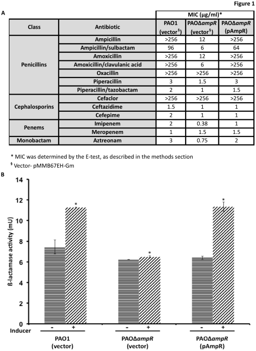
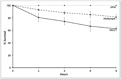
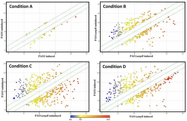
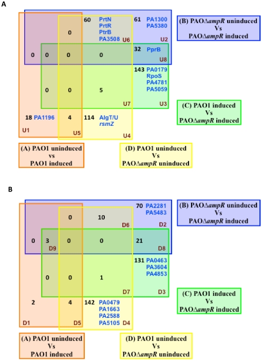
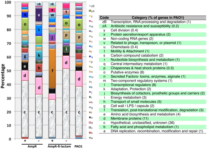
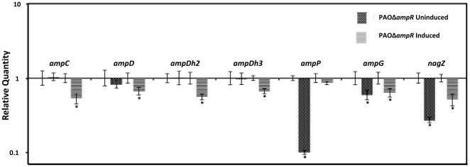
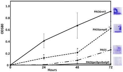
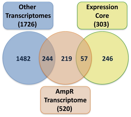
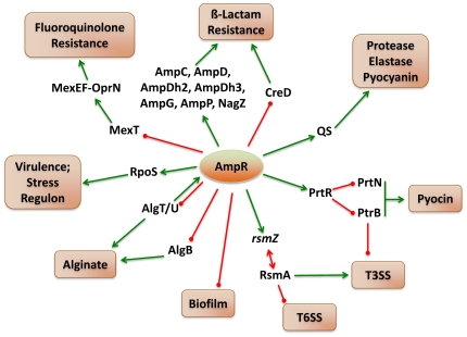
In Enterobacteriaceae, the transcriptional regulator AmpR, a member of the LysR family, regulates the expression of a chromosomal β-lactamase AmpC. The regulatory repertoire of AmpR is broader in Pseudomonas aeruginosa, an opportunistic pathogen responsible for numerous acute and chronic infections including cystic fibrosis. In addition to regulating ampC, P. aeruginosa AmpR regulates the sigma factor AlgT/U and production of some quorum sensing (QS)-regulated virulence factors. In order to better understand the ampR regulon, we compared the transcriptional profile generated using DNA microarrays of the prototypic P. aeruginosa PAO1 strain with its isogenic ampR deletion mutant, PAOΔampR. Transcriptome analysis demonstrates that the AmpR regulon is much more extensive than previously thought, with the deletion of ampR influencing the differential expression of over 500 genes. In addition to regulating resistance to β-lactam antibiotics via AmpC, AmpR also regulates non-β-lactam antibiotic resistance by modulating the MexEF-OprN efflux pump. Other virulence mechanisms including biofilm formation and QS-regulated acute virulence factors are AmpR-regulated. Real-time PCR and phenotypic assays confirmed the microarray data. Further, using a Caenorhabditis elegans model, we demonstrate that a functional AmpR is required for P. aeruginosa pathogenicity. AmpR, a member of the core genome, also regulates genes in the regions of genome plasticity that are acquired by horizontal gene transfer. Further, we show differential regulation of other transcriptional regulators and sigma factors by AmpR, accounting for the extensive AmpR regulon. The data demonstrates that AmpR functions as a global regulator in P. aeruginosa and is a positive regulator of acute virulence while negatively regulating biofilm formation, a chronic infection phenotype. Unraveling this complex regulatory circuit will provide a better understanding of the bacterial response to antibiotics and how the organism coordinately regulates a myriad of virulence factors in response to antibiotic exposure.


Conflict of interest statement

Competing Interests: The authors have declared that no competing interests exist.

Figures

Figure 1
Figure 1. Antibiotic resistance profile of PAOΔampR.
A clean in-frame deletion of ampR was generated in P. aeruginosa PAO1 as described in the methods section to generate PAOΔampR. Figure 1A shows the resistance profile of the strains to the four major classes of β-lactam antibiotics. Representative data from three different biological replicate trials are shown. The amount of β-lactamase produced was quantified (Fig. 1B) in the presence (+) and absence (−) of sub-MIC concentration of a β-lactam inducer.
Figure 2
Figure 2. Effect of ampR deletion on pathogenicity to C. elegans.
The fast killing assay was used to test the effect of loss of ampR on the C. elegans killing ability of PAO1. p-value<0.05 at all time points.
Figure 3
Figure 3. Scatter plots of significantly regulated genes.
Only genes that showed significant (p≤0.01) differential regulation under the various conditions are depicted as colored squares. The colors represent the extent of gene expression from low (blue) to high (red) in either condition, as depicted in the color scale. The two outer green diagonal lines in each plot represent the two-fold cutoff. Each sub-plot depicts the differential gene expression between two strains/conditions (shown along the plot axes): Condition A- PAO1 uninduced vs. PAO1 induced; Condition B- PAOΔampR uninduced vs. PAOΔampR induced; Condition C- PAO1 uninduced vs. PAOΔampR uninduced; Condition D- PAO1 induced vs. PAOΔampR induced.
Figure 4
Figure 4. Venn diagram of the differentially regulated genes.
Distribution of significantly (p≤0.01) differentially regulated (>2-fold) genes in PAO1 and PAOΔampR with (induced) and without (uninduced) β-lactam treatment showing upregulated (Fig. 4A) and downregulated (Fig. 4B) genes. The transcriptional regulators, sigma factors and small RNAs in each group are identified either by their gene names or PA numbers. Annotations are from the Pseudomonas Genome database .
Figure 5
Figure 5. Enrichment of functional categories.
Functional categorization of the AmpR-dependent and AmpR-β-lactam dependent genes was performed to identify enrichment of specific classes of genes relative to their distribution in PAO1. ‘+’ and ‘−’ refers to upregulation and downregulation of genes, respectively in either an AmpR-dependent or AmpR-β-lactam-dependent manner. The most differentially regulated categories are labeled in the figure using the corresponding code, mentioned in the figure table. Functional categories, and their codes and percentages in PAO1 are from the Pseudomonas Genome database .
Figure 6
Figure 6. AmpR binding site analysis.
The putative AmpR binding site was refined using IEM algorithm and used to search the upstream promoter elements of AmpR-regulated and AmpR-β-lactam regulated genes (listed in Tables S3, S4) in PAO1 and other P. aeruginosa strains. DOOR database , was used to identify the operons. The RSAT tool was used to identify PAO1 promoter sequences containing the identified AmpR- and AmpR-β-lactam-regulated genes. The output is represented using WebLogo for the AmpR-dependent genes from IEM (A) and RSA (B), and AmpR-β-lactam dependent genes from IEM (C) and RSA (D).
Figure 7
Figure 7. Expression of the amp genes in PAOΔampR.
RNA was isolated from cells exposed to β-lactam antibiotic, reverse transcribed to cDNA and tested in triplicate by qPCR with gene-specific primers, as described in the text. The expression of ampC (β-lactamase), ampD, ampDh2, and ampDh3 (amidases), ampG and ampP (permeases), and nagZ (glycoside hydrolase) were tested in the ampR mutant relative to PAO1. Values have been normalized to expression in PAO1 under the same conditions (log10 RQ = 1) and bars above or below the line represent up- and down-regulation, respectively. * p value<0.05.
Figure 8
Figure 8. AmpR affects biofilm formation.
AmpR regulates many genes involved in biofilm formation (Table S6) that is reflected in the tube biofilm assay monitored for 72 hours. The strains tested were wild type PAO1, PAOΔampR, PAOΔretS (positive control), and PAOΔpelΔpslΔalgD (negative control). The inset, taken at 72 hours, demonstrates the superior biofilm formation capacity of PAOΔampR compared to PAO1. p-values comparing PAOΔampR with PAO1: 24 hrs- 0.002; 48 hrs- 0.03; 72 hrs- 0.007.
Figure 9
Figure 9. Regulation of secretion genes by AmpR.
Fold changes (FC) as determined by microarray experiments in the absence of β-lactam stress of the Type VI secretion (▴, HSI-I) and Type III secretion (▪) genes in the ampR mutant strain, normalized to expression in the wild-type strain PAO1. Gene annotations are from the Pseudomonas Genome database .
Figure 10
Figure 10. Gene expression in PAOΔampR at 40 minutes and 2 hrs post-β-lactam exposure.
RNA was isolated from PAO1 and PAOΔampR cells exposed to β-lactams for either 40 minutes or 2 hours and reverse transcribed to cDNA. The expression of the sigma factor rpoS, lecA and lecB (galactophilic lectin genes known to be RpoS-regulated), and mexT (MexEF-OprN efflux pump regulator that is not RpoS-regulated) were tested by qPCR with gene-specific primers, as described in the text. Values have been normalized to expression in PAO1 under the respective conditions. ** p-value<0.006; *** p-value 0.0002 as determined by unpaired t test.
Figure 11
Figure 11. Comparison of the AmpR transcriptome with other transcriptomes.
AmpR-dependent and AmpR-β-lactam-dependent genes were compared with the 303 genes of the expression core and the 1726 condition-specific genes identified previously as part of a meta-analysis of 18 P. aeruginosa transcriptomes .
Figure 12
Figure 12. AmpR is a master regulator of gene expression in P. aeruginosa PAO1.
AmpR positively regulates resistance to β-lactam antibiotics by upregulating expression of the amp genes, nagZ and downregulating creD. In addition, AmpR affects fluoroquinolone resistance by negatively regulating expression of mexT, the positive regulator of the MexEF-OprN efflux pump. Expression of the virulence and stress response sigma factor, RpoS and QS-regulated acute virulence factors is downregulated in PAOΔampR, indicating positive AmpR regulation. AmpR also negatively regulates biofilm formation via an unknown mechanism. AmpR modulates levels of the small RNA rsmZ, whose levels are lower in PAOΔampR with a corresponding enhanced expression of RsmA. Downregulation of some of the T3SS genes in the ampR mutant is possibly by regulating ptrB expression, via PrtR. Further, two major regulators of the alginate biosynthetic pathway, AlgT/U and AlgB are negatively regulated by AmpR, thereby potentially also regulating alginate production. Whether these AmpR interactions are direct or indirect needs to be investigated.

References

    1. Obritsch MD, Fish DN, MacLaren R, Jung R. National surveillance of antimicrobial resistance in Pseudomonas aeruginosa isolates obtained from intensive care unit patients from 1993 to 2002. Antimicrob Agents Chemother. 2004;48:4606–4610. - PMC - PubMed
    1. Bouza E, Burillo A, Munoz P. Catheter-related infections: diagnosis and intravascular treatment. Clin Microbiol Infect. 2002;8:265–274. - PubMed
    1. Gallagher PG, Watanakunakorn C. Pseudomonas bacteremia in a community teaching hospital, 1980–1984. Rev Infect Dis. 1989;11:846–852. - PubMed
    1. Collin BA, Leather HL, Wingard JR, Ramphal R. Evolution, incidence, and susceptibility of bacterial bloodstream isolates from 519 bone marrow transplant patients. Clin Infect Dis. 2001;33:947–953. - PubMed
    1. Davies JC. Pseudomonas aeruginosa in cystic fibrosis: pathogenesis and persistence. Paediatr Respir Rev. 2002;3:128–134. - PubMed

Publication types

MeSH terms