. 2012;7(3):e34570.
doi: 10.1371/journal.pone.0034570. Epub 2012 Mar 30.

Could a B-1 cell derived phagocyte "be one" of the peritoneal macrophages during LPS-driven inflammation?

Affiliations

Could a B-1 cell derived phagocyte "be one" of the peritoneal macrophages during LPS-driven inflammation?

Ana Flavia Popi et al. PLoS One. 2012.

Abstract

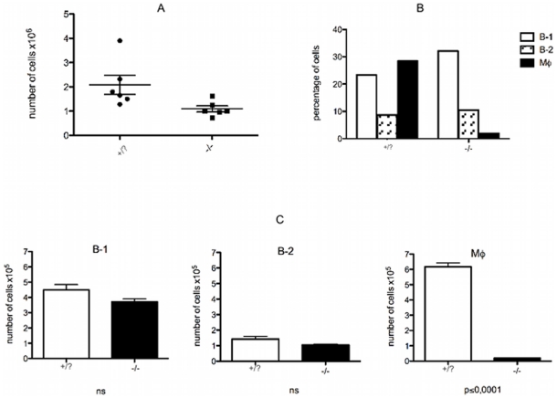
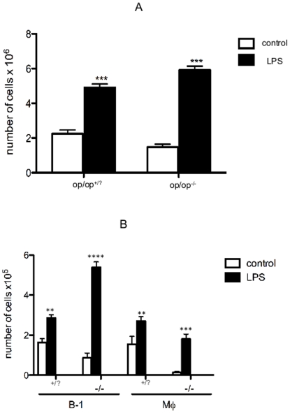
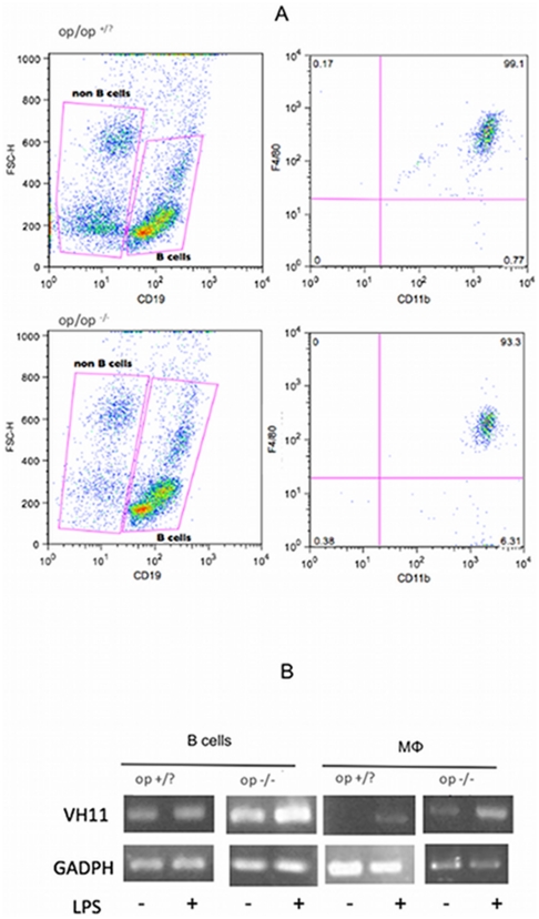
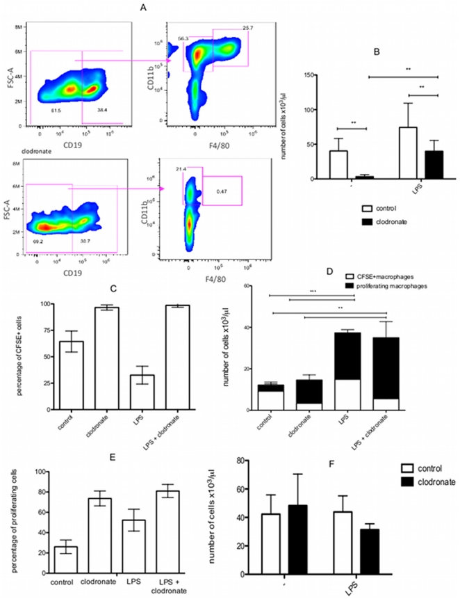
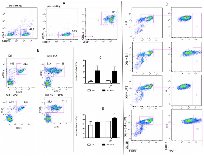
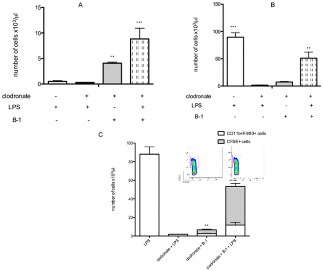
The inflammatory response is driven by signals that recruit and elicit immune cells to areas of tissue damage or infection. The concept of a mononuclear phagocyte system postulates that monocytes circulating in the bloodstream are recruited to inflamed tissues where they give rise to macrophages. A recent publication demonstrated that the large increase in the macrophages observed during infection was the result of the multiplication of these cells rather than the recruitment of blood monocytes. We demonstrated previously that B-1 cells undergo differentiation to acquire a mononuclear phagocyte phenotype in vitro (B-1CDP), and we propose that B-1 cells could be an alternative origin for peritoneal macrophages. A number of recent studies that describe the phagocytic and microbicidal activity of B-1 cells in vitro and in vivo support this hypothesis. Based on these findings, we further investigated the differentiation of B-1 cells into phagocytes in vivo in response to LPS-induced inflammation. Therefore, we investigated the role of B-1 cells in the composition of the peritoneal macrophage population after LPS stimulation using osteopetrotic mice, BALB/Xid mice and the depletion of monocytes/macrophages by clodronate treatment. We show that peritoneal macrophages appear in op/op((-/-)) mice after LPS stimulation and exhibit the same Ig gene rearrangement (VH11) that is often found in B-1 cells. These results strongly suggest that op/op((-/-)) peritoneal "macrophages" are B-1CDP. Similarly, the LPS-induced increase in the macrophage population was observed even following monocyte/macrophage depletion by clodronate. After monocyte/macrophage depletion by clodronate, LPS-elicited macrophages were observed in BALB/Xid mice only following the transfer of B-1 cells. Based on these data, we confirmed that B-1 cell differentiation into phagocytes also occurs in vivo. In conclusion, the results strongly suggest that B-1 cell derived phagocytes are a component of the LPS-elicited peritoneal macrophage population.


Conflict of interest statement

Competing Interests: The authors have declared that no competing interests exist.

Figures

Figure 1
Figure 1. Characterization of peritoneal cell subpopulations in the peritoneal cavity of op/op(−/−) mice.
(A) The number of cells in the peritoneal cavity of op/op(−/−) mice is significantly reduced when compared to the littermates. Peritoneal cells were collected from op/op(−/−) mice and their normal littermates (op/op(+/?)), and the cells were counted (p<0.01). (B) Percentage of B-1 cells, B-2 cells and macrophages in the peritoneal cavity of op/op(−/−) mice and littermates (op/op(+/?)). (C) Absolute number of B-1 cells (ns), B-2 cells (ns) and macrophages (Mϕ - p<0.0001). Cell populations were determined by flow analysis based on the following surface marker expression: B-1 cells (CD23CD19+CD11b+), Mϕ (CD11b+F4/80+CD19), and B-2 (CD23+CD19+CD11b). Graphs are representative of two independent experiments using 6 mice per group per experiment.
Figure 2
Figure 2. LPS increases the number of B-1 cells and macrophages in the peritoneal cavity of op/op(−/−) mice.
(A) Number of peritoneal cells collected and counted individually from 6 mice (op/op(−/−)) and their littermates (op/op(+/?)) in control or LPS-treated groups. (B) Absolute number of B-1 cells and macrophages in untreated control (□) or LPS stimulated (▪) littermates op/op(+/?) and op/op(−/−) mice. Cell populations were determined by flow analysis based on surface marker expression: B-1 cells (CD23CD19+CD11b+) and Mϕ (CD11b+F4/80+CD19). Flow cytometric analysis showed significantly increased percentages of B-1 cells in both mice lineages after LPS stimulation when compared to untreated mice. LPS also increased the number of macrophage cells in both groups of mice. Significant differences between the sample means are indicated as follows: **p<0.05, ***p<0.01, and ****p<0.001. The results are representative of two independent experiments.
Figure 3
Figure 3. Elicited peritoneal “macrophages” from op/op(−/−) mice exhibit Ig gene rearrangements.
A) Flow cytometry analysis of total peritoneal cells to distinguish B cells (CD19+) and non-B cells (CD19). After cell purification, the CD19+ cells and the CD19 cells were collected separately. The right dot plots show that non-B cells are F4/80+CD11b+ and are thus characterized as macrophages. B) Analysis of heavy chain variable gene VH11 expression by peritoneal B cells and macrophages from unstimulated or LPS-stimulated op/op(+/?) and op/op(−/−) mice. The PCR products were visualized in agarose gels by ethidium bromide staining. The amount of cDNA input was normalized by analyzing the control GADPH transcripts. The results are representative of three independent experiments using 8 mice per group per experiment.
Figure 4
Figure 4. Clodronate treatment did not impede the proliferation of resident peritoneal “macrophages” from BALB/c mice treated with LPS.
A) Peritoneal cells were collected from untreated and clodronate-treated mice 3 days after the last clodronate injection. Non-B cells were selected based on the total peritoneal CD19 cells, and the CD11b+F4/80+ cells were considered macrophages. This CD11b+F4/80+ cell population was not observed after clodronate treatment. B) Absolute number of peritoneal macrophages (CD19CD11b+F4/80+) from non-clodronate treated (control group □) or clodronate-treated (▪) BALB/c mice after LPS or saline (−) injection. C) Percentage of CFSE+ cells present in the peritoneal macrophage population (CD19CD11b+F4/80+) in BALB/c mice subjected to different treatments: control (without clodronate+saline injection); clodronate (clodronate+saline); LPS (without clodronate+LPS injection) and LPS+clodronate (clodronate+LPS). The same group legend was used in figure 4D and 4E. D) Absolute number of CFSE+ macrophages (CFSE+CD19CD11b+F4/80+) showing high CFSE fluorescence (non-proliferating cells; CFSE+macrophages) and the decay in CFSE fluorescence (proliferating macrophages). E) Percentage of proliferating macrophages (CFSEdim) detected in different treated BALB/c mice. F) Absolute number of peritoneal B-1 cells (CD23CD19+CD11b+) in non-clodronate treated (control group □) or clodronate-treated (▪) BALB/c mice after LPS or saline (−) injection. **p<0.01 and ***p<0.001 when the indicated groups are compared. The results are representative of two or three independent experiments using 5 mice per group per experiment.
Figure 5
Figure 5. LPS induces B-1CDP differentiation and proliferation.
A) Analysis of B-1 cell purification and CFSE+ staining. B-1 cells were obtained from the total peritoneal cells based on the selection of CD19+CD23 cells as described in Figure S1. The first dot plot represents the B-1 cell gate selected to perform cell sorting. The second dot plot illustrates B-1 cells after cell sorting. Subsequently, these cells were subjected to CFSE staining as demonstrated in the last dot plot. B) Analysis of expression of CD19 and CD23 by peritoneal lymphocytes confirms the transfer of B-1 cells to BALB/Xid mice. B-1 cells are almost absent in BALB/Xid and BALB/Xid+LPS mice but are present in significant numbers in BALB/Xid+B-1 and BALB/Xid+B-1+LPS mice, indicating the efficiency of the B-1 cell transfer. C) Absolute number of B-1 cells (CD19+CD23CD11b+) in the peritoneal cavity of BALB/Xid (□) and BALB/Xid+B-1 (▪) after LPS or saline (−) injection. p<0.001 when the LPS-stimulated group is compared to the non-stimulated group. D) Analysis of the presence of B-1CDP (CFSE+ cells) in the peritoneal macrophage population (CD19CD11b+F4/80+) from BALB/Xid, BALB/Xid+B-1, BALB/Xid+LPS and BALB/Xid+B-1+LPS mice. E) Absolute number of peritoneal macrophages (CD19CD11b+F4/80+) from BALB/Xid mice (□) and BALB/Xid+B-1 mice (▪) after LPS or saline (−) injection. * p<0.01 when the indicated group is compared to all groups. The results are representative of two independent experiments using 5 mice per group per experiment.
Figure 6
Figure 6. B-1CDPs are one of the components of the peritoneal macrophage population after LPS stimulation.
A) Absolute number of peritoneal B-1 cells (CD19+CD23CD11b+) detected in BALB/Xid mice submitted to the specified treatment. ** p<0.01 and ***p<0.001 when the indicated groups are compared with the non-marked groups. p<0.001 when the two last groups are compared. B) Absolute number of peritoneal macrophages (CD19CD11b+F4/80+) detected in BALB/Xid mice submitted to the specified treatment. ** p<0.01 and ***p<0.001 when the indicated groups are compared with the non-marked groups. C) Absolute number of macrophages (CD19CD11b+F4/80+) and the number of B-1CDPs (CFSE+ cells) detected in this population from BALB/Xid mice submitted to the specified treatment. **p<0.01 and ****p<0.001 when the CFSE+ cell number of the indicated groups are compared with all other groups. The results are representative of two independent experiments using 4 or 5 mice per group per experiment.

References

    1. van Furth R, Cohn ZA, Hirsch JG, Humphrey JH, Spector WG, et al. The mononuclear phagocyte system: a new classification of macrophages, monocytes, and their precursor cells. Bull World Health Organ. 1972;46:845–852. - PMC - PubMed
    1. Yamashiro S, Takeya M, Kuratsu J, Ushio Y, Takahashi K, et al. Intradermal injection of monocyte chemoattractant protein-1 induces emigration and differentiation of blood monocytes in rat skin. Int Arch Allergy Immunol. 1998;115:15–23. - PubMed
    1. Naito M, Hayashi S, Yoshida H, Nishikawa S, Shultz LD, et al. Abnormal differentiation of tissue macrophage populations in ‘osteopetrosis’ (op) mice defective in the production of macrophage colony-stimulating factor. Am J Pathol. 1991;139:657–667. - PMC - PubMed
    1. Umeda S, Takahashi K, Shultz LD, Naito M, Takagi K. Effects of macrophage colony-stimulating factor on macrophages and their related cell populations in the osteopetrosis mouse defective in production of functional macrophage colony-stimulating factor protein. Am J Pathol. 1996;149:559–574. - PMC - PubMed
    1. Takahashi K, Naito M, Takeya M. Development and heterogeneity of macrophages and their related cells through their differentiation pathways. Pathol Int. 1996;46:473–485. - PubMed

Publication types

Substances