. 2012 Dec;28(12):1693-702.
doi: 10.1089/AID.2012.0028. Epub 2012 May 4.

CD4 T cell depletion exacerbates acute Mycobacterium tuberculosis while reactivation of latent infection is dependent on severity of tissue depletion in cynomolgus macaques

Affiliations

CD4 T cell depletion exacerbates acute Mycobacterium tuberculosis while reactivation of latent infection is dependent on severity of tissue depletion in cynomolgus macaques

Philana Ling Lin et al. AIDS Res Hum Retroviruses. 2012 Dec.

Abstract

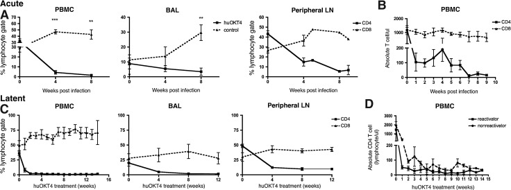
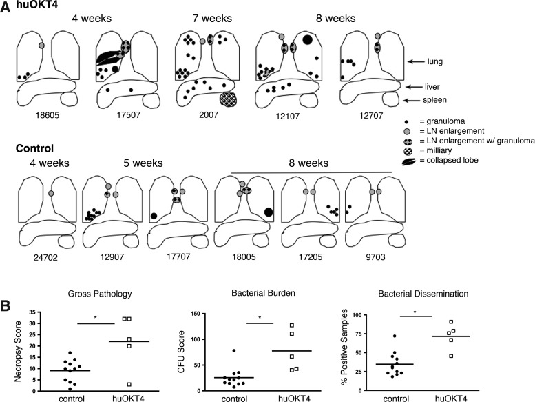
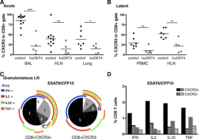
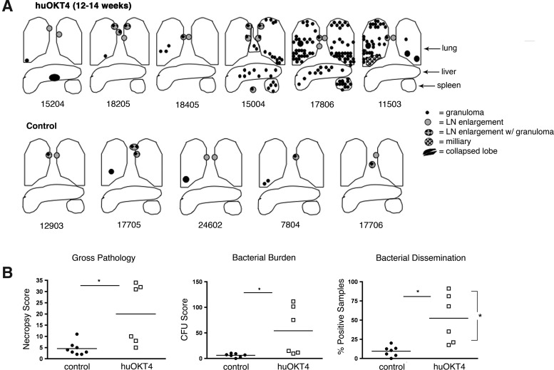
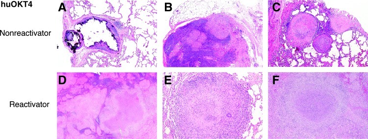
CD4 T cells are believed to be important in protection against Mycobacterium tuberculosis, but the relative contribution to control of initial or latent infection is not known. Antibody-mediated depletion of CD4 T cells in M. tuberculosis-infected cynomolgus macaques was used to study the role of CD4 T cells during acute and latent infection. Anti-CD4 antibody severely reduced levels of CD4 T cells in blood, airways, and lymph nodes. Increased pathology and bacterial burden were observed in CD4-depleted monkeys during the first 8 weeks of infection compared to controls. CD4-depleted monkeys had greater interferon (IFN)-γ expression and altered expression of CD8 T cell activation markers. During latent infection, CD4 depletion resulted in clinical reactivation in only three of six monkeys. Reactivation was associated with lower CD4 T cells in the hilar lymph nodes. During both acute and latent infection, CD4 depletion was associated with reduced percentages of CXCR3(+) expressing CD8 T cells, reported to be involved in T cell recruitment, regulatory function, and effector and memory T cell maturation. CXCR3(+) CD8 T cells from hilar lymph nodes had more mycobacteria-specific cytokine expression and greater coexpression of multiple cytokines compared to CXCR3(-) CD8 T cells. CD4 T cells are required for protection against acute infection but reactivation from latent infection is dependent on the severity of depletion in the draining lymph nodes. CD4 depletion influences CD8 T cell function. This study has important implications for human HIV-M. tuberculosis coinfection.


Figures

FIG. 1.
FIG. 1.
huOKT4A severely depletes CD4 T cells from blood, bronchoalveolar lavage (BAL), and peripheral lymph node (LN) during acute and latent Mycobacterium tuberculosis (Mtb) infection. (A) Frequency of CD4 T cells from peripheral blood mononuclear cells (PBMCs), BAL, and peripheral LN in huOKT4-treated (n=5) and control monkeys (n=7) during acute infection (**p<0.01, ***p<0.001, Student's t-test). (B) huOKT4-depleted CD4 T cells in blood to <200 cells/(l. (C) huOKT4 severely depleted CD4 T cells in latently infected animals in PBMCs, BAL, and peripheral LN. (D) No difference in absolute blood CD4 T cell levels between reactivators and nonreactivators (Mann–Whitney analysis). Means are shown with standard error of the mean (SEM).
FIG. 2.
FIG. 2.
huOKT4A exacerbates disease following acute Mtb infection. (A) Gross pathology is illustrated in CD4-depleted monkeys (top panel) and representative acute controls 4–8 weeks postinfection (pi); monkey numbers are below the diagrams. (B) huOKT4A-treated monkeys had more pathology (necropsy score), bacterial burden (CFU score), and dissemination (% positive samples) compared to control monkeys (*p<0.05, Mann–Whitney). Each point on the figure represents a monkey. Median values are shown.
FIG. 3.
FIG. 3.
CD4 depletion results in fewer CXCR3+CD8+ T cells and these cells produce more cytokines in active tuberculosis. Significantly reduced frequencies of CXCR3+CD8 T cells were observed in huOKT4-treated monkeys during (A) acute infection and (B) latent infection (*p<0.05, **p<0.01, ***p<0.001; Student's t-test). Each circle represents an animal. (C, D) Cytokine production from CXCR3+CD8+ T cells from granulomatous hilar LN (HLN) was increased compared to CXCR3CD8+ T cells in active tuberculosis controls, and was higher than in nongranulomatous lymph node (LN). (C) Numbers in pies represent the number of cytokines coexpressed in CD8 T cells. The colored arcs depict the specific cytokine produced. (D) Granulomatous hilar LN (black); nongranulomatous LN (gray); solid bars CXCR3+, checkered bars CXCR3 CD8 T cells. Median values are shown.
FIG. 4.
FIG. 4.
huOKT4A treatment reactivates latent infection in three of six monkeys. (A) Diagram depictions of gross pathology of huOKT4A-treated and control latent monkeys; monkey numbers are below the diagrams. (B) huOKT4A monkeys had higher gross pathology (necropsy score), bacterial burden (CFU score), and dissemination (% positive samples) compared to latent controls. Reactivators had greater dissemination than nonreactivators (*p<0.05, Mann–Whitney). Median values are shown.
FIG. 5.
FIG. 5.
huOKT4A-treated latent monkeys had histopathologic signs of latent lesions and reactivation. (A, B, C) huOKT4A-treated latent monkeys that did not reactivate had classic mineralized granulomas associated with latent infection (A) (5×). Microscopic findings suggestive of early reactivation in 18205 included nonnecrotizing granulomas in hilar LN (B) (5×) and nonnecrotizing, solid fibrous, and sclerotic granulomas in lungs (C) (5×). (D, E) huOKT4A-treated monkeys that reactivated had histology consistent with active/reactivated tuberculosis: multifocal areas of caseous and nonnecrotizing granulomas effacing hilar LN (D) (2×) and large caseous granulomas in lungs (E) (10×). (F) Extrapulmonary disease included caseous granulomas in spleen (5×) and liver (not shown). H&E staining.

References

    1. Verver S, et al. Transmission of tuberculosis in a high incidence urban community in South Africa. Int J Epidemiol. 2004;33(2):351–357. - PubMed
    1. Selwyn PA, et al. Clinical manifestations and predictors of disease progression in drug users with human immunodeficiency virus infection. N Engl J Med. 1992;327(24):1697–1703. - PubMed
    1. Lawn SD, et al. Impact of HIV infection on the epidemiology of tuberculosis in a peri-urban community in South Africa: The need for age-specific interventions. Clin Infect Dis. 2006;42(7):1040–1047. - PubMed
    1. Lawn SD, et al. Short-term and long-term risk of tuberculosis associated with CD4 cell recovery during antiretroviral therapy in South Africa. AIDS. 2009;23(13):1717–1725. - PMC - PubMed
    1. Barnes PF, et al. Tuberculosis in patients with HIV infection. Infect Dis Clin North Am. 2002;16(1):107–126. - PubMed

Publication types

MeSH terms