Review
. 2012 May;22(5):241-9.
doi: 10.1016/j.tcb.2012.02.007. Epub 2012 Apr 3.

Acentrosomal spindle assembly and chromosome segregation during oocyte meiosis

Affiliations
Review

Acentrosomal spindle assembly and chromosome segregation during oocyte meiosis

Julien Dumont et al. Trends Cell Biol. 2012 May.

Abstract

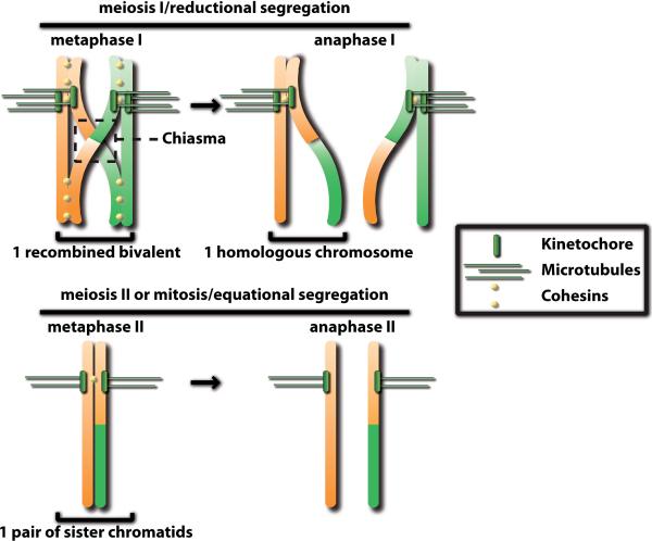
The ability to reproduce relies in most eukaryotes on specialized cells called gametes. Gametes are formed by the process of meiosis in which, after a single round of replication, two successive cell divisions reduce the ploidy of the genome. Fusion of gametes at fertilization reconstitutes diploidy. In most animal species, chromosome segregation during female meiosis occurs on spindles assembled in the absence of the major microtubule-organizing center, the centrosome. In mammals, oocyte meiosis is error prone and underlies most birth aneuploidies. Here, we review recent work on acentrosomal spindle formation and chromosome alignment/separation during oocyte meiosis in different animal models.


Figures

Figure 1
Figure 1. Chromosome segregation during meiosis
Recombined homologous chromosomes are held together by sister chromatid cohesion after exchange of genetic material during meiotic recombination at the chiasma. During anaphase I, dissolution of chromosome arm cohesion leads to reductional segregation of homologs. Sister chromatids are equationally segregated following dissolution of centromeric cohesion during anaphase II.
Figure 2
Figure 2. Models of centrosomal and acentrosomal spindle assembly
Centrosomal spindles assemble in somatic cells, in spermatocytes and in echinoderm oocytes. Microtubules nucleated from the centrosomes (yellow) oscillate between phases of growth and shrinkage `searching' for chromosomes. End-on contact with a kinetochore results in `capture' of a chromosome and stabilization of the microtubule to form a kinetochore fiber (orange). In mouse oocytes, multiple MTOCs (yellow) assemble microtubule aster-like structures in the vicinity of chromosomes that subsequently are organized into a bipolar spindle. Kinetochore fibers are periodically destabilized and re-established to maintain oscillating chromosomes at the spindle equator during the extended prometaphase. In Drosophila oocytes, microtubule asters first assemble at NEBD away from chromosomes in the absence of discrete PCM-containing MTOCs. These acentrosomal asters are subsequently remodeled and incorporated into the forming spindle, which is assembled by sorting and focusing of microtubule (−)-ends at the spindle poles. In cell-free Xenopus extracts, microtubules are nucleated in the vicinity of chromatin-coated beads in random orientation and are subsequently sorted into an antiparallel microtubule array that surrounds chromosomes. Microtubule (−)-ends are focused away from chromosomes to form the spindle poles.
Figure 3
Figure 3. Chromatin-dependent pathways of microtubule assembly
During M-Phase, spindle assembly factors (SAFs) are sequestered by Importins that bind to their NLS. A gradient of active Ran (RanGTP; orange) centered on chromosomes locally releases spindle assembly factors (SAFs) from the inhibitory effect of Importins. In parallel, Aurora B, which is activated by clustering at the inner centromere or on microtubules, phosphorylates and inhibits the microtubule depolymerase MCAK, and Oncoprotein 18 (OP18) to reduce its ability to sequester tubulin heterodimers. The Ran and CPC-dependent pathways create a locally favorable environment for microtubule and spindle assembly.
Figure 4
Figure 4. Mechanisms of anaphase chromosome physical separation
During anaphase A, shortening of kinetochore-attached microtubules moves chromosomes toward the spindle poles without increase in the pole-to-pole distance. Anaphase B involves separation of the spindle poles while chromosome-to-pole distance remains constant. Spindle pole separation relies on sliding of antiparallel interpolar microtubules and/or cortical pulling forces exerted on astral microtubules. Both anaphase A and B chromosome physical separation requires functional kinetochores with end-on attached microtubules. Central spindle-driven anaphase relies on microtubule assembly between the separating chromosomes and is kinetochore-independent. The nature of the link between microtubules and chromosomes, and whether microtubule assembly and/or sliding promote chromosome physical separation in this mechanism are still unclear.

References

    1. Hassold T, Hunt P. Maternal age and chromosomally abnormal pregnancies: what we know and what we wish we knew. Curr Opin Pediatr. 2009;21:703–708. - PMC - PubMed
    1. Nigg EA, Raff JW. Centrioles, centrosomes, and cilia in health and disease. Cell. 2009;139:663–678. - PubMed
    1. Manandhar G, et al. Centrosome reduction during gametogenesis and its significance. Biol Reprod. 2005;72:2–13. - PubMed
    1. Szöllösi D, et al. Absence of centrioles in the first and second meiotic spindles of mouse oocytes. J Cell Sci. 1972;11:521–541. - PubMed
    1. Dumont J, Brunet S. Meiotic Spindle Assembly and Chromosome Segregation in Oocytes. In: Verlhac MH, Villeneuve AM, editors. Oogenesis: The Universal Process. Wiley-Blackwell; 2010. pp. 269–290.

Publication types