Review
. 2012 May;37(5):179-88.
doi: 10.1016/j.tibs.2012.02.009. Epub 2012 Apr 3.

The spliceosome: a flexible, reversible macromolecular machine

Affiliations
Review

The spliceosome: a flexible, reversible macromolecular machine

Aaron A Hoskins et al. Trends Biochem Sci. 2012 May.

Abstract

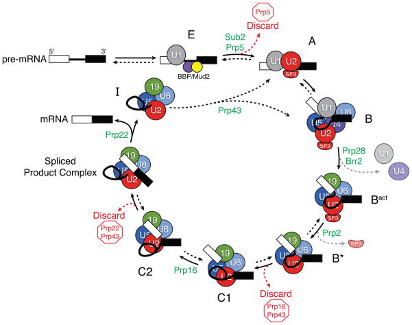
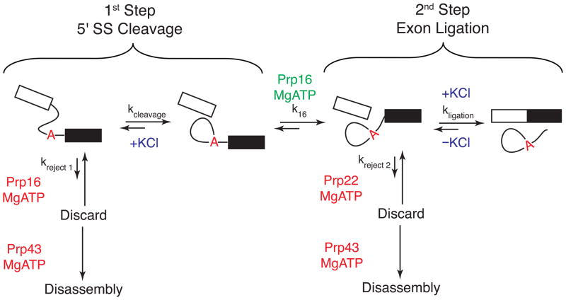
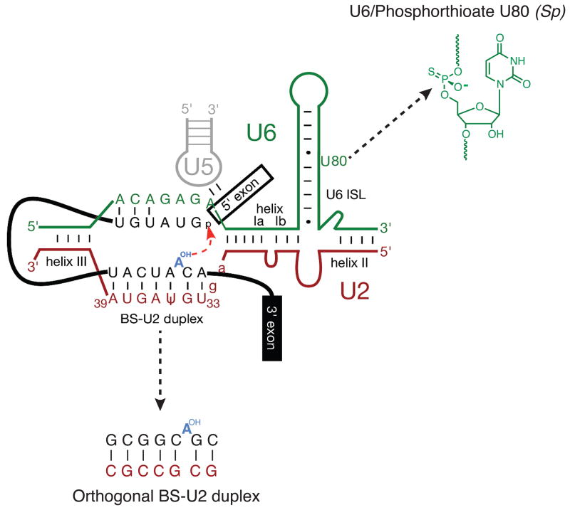
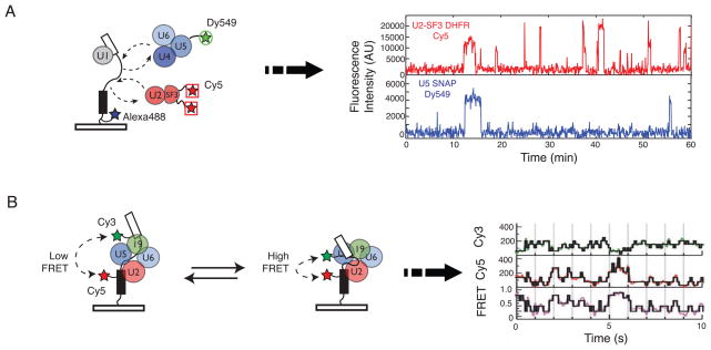
With more than a hundred individual RNA and protein parts and a highly dynamic assembly and disassembly pathway, the spliceosome is arguably the most complicated macromolecular machine in the eukaryotic cell. This complexity has made kinetic and mechanistic analysis of splicing incredibly challenging. Yet, recent technological advances are now providing tools for understanding this process in much greater detail. Ranging from genome-wide analyses of splicing and creation of an orthogonal spliceosome in vivo, to purification of active spliceosomes and observation of single molecules in vitro, such new experimental approaches are yielding significant insight into the inner workings of this remarkable machine. These experiments are rewriting the textbooks, with a new picture emerging of a dynamic, malleable machine heavily influenced by the identity of its pre-mRNA substrate.


Figures

Figure 1
Figure 1
A model for step-wise spliceosome assembly and catalysis. Specific spliceosomal complexes (E, A, B, and others) are identified according to the human nomenclature. Spliceosome assembly initiates by binding of the U1 snRNP to the 5′ SS and proteins [branch point bridging protein (BBP) and Mud2] to the branch site in E (early) complex. In an ATP-dependent reaction, U2 displaces BBP/Mud2 and binds to the branch site in A complex. B complex then forms by addition of the U4/U6.U5 tri-snRNP. Subsequent to assembly of B complex, catalytic activation requires several additional rearrangements. These include departure of U1 and U4 to form Bact complex (grey arrow); formation of catalytic structures between the pre-mRNA, U2, and U6 snRNAs; and destabilization of several U2 snRNP proteins (the SF3 complex, grey arrow) from the rest of the machinery to form B* complex [8,12,13]. C1 complex is formed after 5′ SS cleavage. For exon ligation, the spliceosome undergoes a conformational change into C2 complex. After the two chemical steps of splicing are complete, the spliceosome enters a disassembly and recycling pathway in which the spliced exons are released and the post-spliceosomal intron product complex (I) is disrupted. Multiple steps in the pathway are promoted by the presence of DExD/H-box ATPases (green). Some of these ATPases have also been implicated in fidelity checkpoints and control the use of discard pathways (red arrows and type) that prevent splicing. As discussed in the text, many of the steps in the pathway have now been shown to be reversible (double arrows), while others have not (single arrows).
Figure 2
Figure 2
The chemical steps of pre-mRNA splicing and kinetic proofreading. Splicing proceeds through two sequential transesterification reactions: 5′ SS cleavage and exon ligation. These reactions may be catalyzed by alternate conformations of the spliceosome according to the ‘two state’ model for spliceosomal catalysis, and ATP hydrolysis by Prp16 promotes interchange between the two states (k16). As in Figure 1, ATPase functions that promote splicing during these steps are shown in green and those that promote discard pathways are shown in red. The forward or reverse chemical reactions can also be promoted by the presence or absence of KCl (blue) [34]. On a substrate with a high probability of generating mRNA, the forward reaction pathways are favored (large arrows) while the reverse reactions and rejection pathways are disfavored (small arrows). On a poor splicing substrate (or in a malformed spliceosome), the rates of the forward reactions (kcleavage, k16, and kligation) may be slower than the reactions controlling entrance to the discard pathways (kreject 1 and kreject 2). In this case, Prp43 irreversibly disassembles the spliceosome. This represents a fidelity mechanism for splicing and a version of kinetic proofreading [25].
Figure 3
Figure 3
A model for the spliceosomal B* complex active site showing juxtaposition of several important groups during catalysis. In the work of Koodathingal et al., a site-specific phosphorthioate at position U80 in the U6 snRNA was used to stall spliceosomes prior to exon ligation [24]. Smith et al. created an orthogonal yeast spliceosome by mutating the branch site (BS)-U2 duplex [36]. This figure originated from reference [36] and is used with permission.
Figure 4
Figure 4
Schematic for single molecule fluorescence analysis of splicing. (a) Hoskins et al. tethered fluorescent pre-mRNAs labeled with Alexa488 to a glass surface and then monitored how fluorescent spliceosome subcomplexes (e.g. U2-SF3 and U4/U6.U5 tri-snRNP) associated with the pre-mRNA. Fluctuations in fluorescence intensity indicated that the subcomplexes can associate multiple times with a given pre-mRNA. (b) By incorporating a FRET donor (Cy3) and acceptor (Cy5) pair into a surface-tethered pre-mRNA, Abelson et al. were able to show that pre-mRNA conformation reversibly fluctuated during splicing. These conformational changes are shown by changes in the FRET signal of single pre-mRNA molecules. Data in (a) and (b) are from references [45] and [49], respectively, and used with the authors’ permission.
Figure 5
Figure 5
Microarray analysis of pre-mRNA splicing in yeast and the effects of mutations in core spliceosomal proteins. Each pre-mRNA in yeast (such as the subset of 12 shown here) can respond uniquely to mutations in core splicing factors. These responses can be identified by either increases (yellow bands) or decreases (blue bands) in pre-mRNA abundance relative to a wild type strain. This indicates that pre-mRNA identity can strongly influence multiple steps in splicing. Data are from reference [53] and used with the authors’ permission.

References

    1. Nilsen TW, Graveley BR. Expansion of the eukaryotic proteome by alternative splicing. Nature. 2010;463:457–463. - PMC - PubMed
    1. Scherer S. Guide to the Human Genome. 1. Cold Spring Harbor; 2010.
    1. Lim LP, Burge CB. A computational analysis of sequence features involved in recognition of short introns. Proc Natl Acad Sci USA. 2001;98:11193–11198. - PMC - PubMed
    1. Roca X, Krainer AR. Recognition of atypical 5′ splice sites by shifted base-pairing to U1 snRNA. Nat Struct Mol Biol. 2009;16:176–182. - PMC - PubMed
    1. Wahl MC, et al. The spliceosome: design principles of a dynamic RNP machine. Cell. 2009;136:701–718. - PubMed

Publication types