Review
. 2012 Jun;28(6):267-75.
doi: 10.1016/j.tig.2012.03.001. Epub 2012 Apr 3.

Genetics of atherosclerosis

Affiliations
Review

Genetics of atherosclerosis

Aldons J Lusis. Trends Genet. 2012 Jun.

Abstract

Genome-wide association studies (GWAS) from the past several years have provided the first unbiased evidence of the genes contributing to common cardiovascular disease traits in European and some Asian populations. The results not only confirmed the importance of prior knowledge, such as the central role of lipoproteins, but also revealed that there is still much to learn about the underlying mechanisms of this disease, as most of the associated genes do not appear to be involved in pathways previously connected to atherosclerosis. In this review, I focus on the common forms of the disease and look at both human and animal model studies. I summarize what was known before GWAS, highlight how the field has been changed by GWAS, and discuss future considerations, such as the limitations of GWAS and strategies that may lead to a more complete, mechanistic understanding of atherosclerosis.


Figures

Figure 1
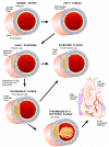
Figure 1. Stages of coronary artery disease
Large arteries are bounded on the luminal side by a monolayer of endothelial cells that form tight junctions, insulating the vessel wall from the blood. Underlying that is a region termed the intima, that consists largely of proteoglycans and collagen. This is followed by layers of smooth muscle cells, and then an outer region, the adventitia, consisting of fibrous elements and fibroblasts. The first stages of atherosclerosis are characterized by the accumulation of lipoproteins, particularly low density lipoproteins (LDL), in the intimal region. In response to the lipoprotein accumulation, monocytes, and subsequently, lymphocytes, infiltrate the vessel wall. The monocytes differentiate to macrophages which then take up the lipoproteins to give rise to cholesterol-engorged “foam cells”, a hallmark of early atherogenesis. Some of these foam cells eventually die, resulting in a necrotic core of cholesterol and cellular debris. This is accompanied by the migration and proliferation of smooth muscle cells, which form a fibrous cap that overlies the necrotic core. The most common cause of a myocardial infarction (MI) is the rupture of an atherosclerotic lesion, exposing tissue factor and leading to the formation of a thrombus that blocks the flow of blood. In general, a thick fibrous cap appears to protect against such rupture (from )
Figure 2
Figure 2. GWAS of CAD in >100,000 Europeans
The Y axis shows the significance of the SNP association with CAD (as the negative of the log10 or the P value), while the X axis shows the location of the SNP across the genome, with Chromosomes shown in alternate colors. The most likely candidate genes at the identified loci are indicated using gene symbols and the floating horizontal line represents genome-wide significance using a Bonferroni adjustment. The names of the genes, in alphabetical order, are as follows: ABO, ABO blood group (transferase A, alpha 1-3-N-acetylgalactosaminyl-transferase B, alpha 1-3-galactosyltransferace); ADAMTS7, ADAM metallopeptidase with thrombospondin type motif 1; ANKS1A, (ankyrin repeat and sterile alpha motif domain containing A1; APOA5-A4-C3-A1, Apolipoprotein A5, A4, C3, A1 gene cluster; CDKN2A, cyclin-dependent kinase inhibitor 2A; CDKN2B, cyclin-dependent kinase inhibitor 2B; COL4A1, collagen, type IV, alpha 1; COL4A2, collagen, type IV, alpha 2; CNNM2, cyclin M2; CXCL12, chemokine (C-X-C motif) ligand 12; CYP17A1, cytochrome P450, family 17, subfamily A, polypeptide 1; CYP46A1, cytochrome P450, family 46, subfamily A, polypeptide 1; HHIPL1, hedgehog interacting protein-like 1; LDLR, low density lipoprotein receptor; LPA, lipoprotein, Lp(a); MIA3, melanoma inhibitory activity family, member 3; MRAS, muscle RAS oncogene homolog; MRPS6, mitochondrial ribosomal protein S6; NT5C2, 5′-nucleotidase, cytosolic II; PCSK9, proprotein convertase subtilisin/kexin type 9; PEMT, phosphatidylethanolamine N-methyltransferase; PHACTR1, phosphatase and actin regulator 1; PPAP2B, phosphatidic acid phosphatase type 2B; RASD1, RAS, dexamethasone-induced 1; SH2B3, SH2B adaptor protein 3; SMG6, smg-6 homolog, nonsense mediated mRNA decay factor, SORT1, sortilin 1; TCF21, transcription factor 21; UBE2Z, ubiquitin-conjugating enzyme E2Z; WDR12, WD repeat domain 12; ZC3HC1, zinc finger, C3HC-type containing 1; ZNF259, zinc finger protein 259. (Reprinted from 25 with permission)
Figure 3
Figure 3. Systems genetics analysis of human endothelial inflammatory response to oxidized phospholipids
Atherosclerosis is initiated by the accumulation of lipoproteins in the vessel wall. The phospholipids and cholesterol can undergo oxidative modification to produce a variety of molecular species, some of which have potent pro-inflammatory activities. In this study, model oxidized phospholipids were used to treat 162 primary human aortic endothelial cell lines, and global transcript levels were determined for treated and control cultures using expression arrays. About 2,000 genes whose expression levels were perturbed by the treatment were used to model a co-expression network using the WGCNA algorithm. A total of 11 subnetwork “modules” of highly connected genes were identified (Panel A) and most were found to be highly enriched for certain known pathways such as cell cycle (shaded green) and the unfolded protein response (shaded purple). Examination of the connections of genes within the modules identified novel pro-inflammatory pathways and regulatory interactions. Shown in Panel (B) is a representation of the purple module (indicated by an arrow) enriched for unfolded protein response genes, with darker colored modules indicating hub nodes, and the length of the edges inversely related to the co-variation. Reprinted from , with permission.

References

    1. Hansson GK. Inflammation, atherosclerosis, and coronary artery disease. N Engl J Med. 2005;352:1685–1695. - PubMed
    1. Libby P, et al. Progress and challenges in translating the biology of atherosclerosis. Nature. 2011;473:317–325. - PubMed
    1. Moore KJ, Tabas I. Macrophages in the pathogenesis of atherosclerosis. Cell. 2011;145:341–355. - PMC - PubMed
    1. Lusis AJ. Atherosclerosis. Nature. 2000;407:233–241. - PMC - PubMed
    1. Maeda N. Development of apolipoprotein E-deficient mice. Arterioscler Thromb Vasc Biol. 2011;31:1957–1962. - PMC - PubMed

Publication types