. 2012 Oct;10(4):341-50.
doi: 10.1007/s12021-012-9147-0.

Volume estimation of the thalamus using freesurfer and stereology: consistency between methods

Affiliations

Volume estimation of the thalamus using freesurfer and stereology: consistency between methods

Simon S Keller et al. Neuroinformatics. 2012 Oct.

Abstract

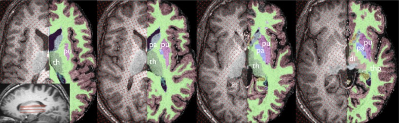
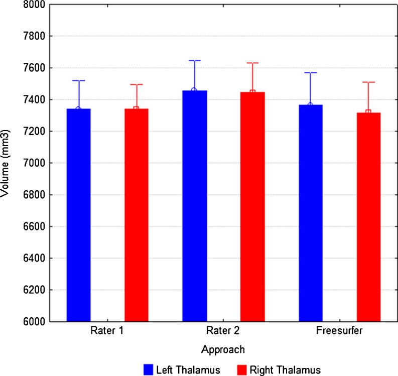
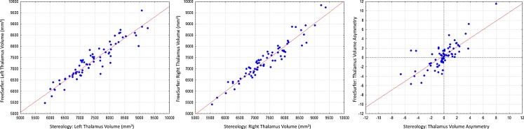
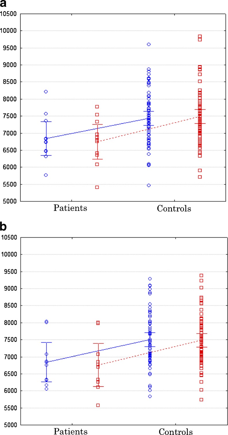
Freely available automated MR image analysis techniques are being increasingly used to investigate neuroanatomical abnormalities in patients with neurological disorders. It is important to assess the specificity and validity of automated measurements of structure volumes with respect to reliable manual methods that rely on human anatomical expertise. The thalamus is widely investigated in many neurological and neuropsychiatric disorders using MRI, but thalamic volumes are notoriously difficult to quantify given the poor between-tissue contrast at the thalamic gray-white matter interface. In the present study we investigated the reliability of automatically determined thalamic volume measurements obtained using FreeSurfer software with respect to a manual stereological technique on 3D T1-weighted MR images obtained from a 3 T MR system. Further to demonstrating impressive consistency between stereological and FreeSurfer volume estimates of the thalamus in healthy subjects and neurological patients, we demonstrate that the extent of agreeability between stereology and FreeSurfer is equal to the agreeability between two human anatomists estimating thalamic volume using stereological methods. Using patients with juvenile myoclonic epilepsy as a model for thalamic atrophy, we also show that both automated and manual methods provide very similar ratios of thalamic volume loss in patients. This work promotes the use of FreeSurfer for reliable estimation of global volume in healthy and diseased thalami.


Figures

Fig. 1
Fig. 1
Stereological and FreeSurfer methods used to estimate thalamic volume shown at approximately the same axial sections. Both methods are shown for the same hemisphere (the FreeSurfer axial sections are flipped to show maximal correspondence between techniques) of the same randomly selected subject. The sagittal MR section illustrates the approximate levels of the axial sections shown. Abbreviations: di, ventral diencephalon (peach); pa, pallidum (violet); put, putamen (lilac); th, thalamus (dark green); thp, pulvinar of the thalamus (dark green)
Fig. 2
Fig. 2
Inter-rater consistency in stereological volume estimation of the left (blue) and right (red) thalamus, and relation to volume estimates obtained from FreeSurfer. Stereological volume estimates are reproducible, and FreeSurfer is entirely consistent with the volumes obtained with both rater one and rater two. Error bars indicate the 95 % confidence intervals
Fig. 3
Fig. 3
Relationship between stereological and FreeSurfer volume estimates of the left a and right b thalamus in the whole study sample. The relationship between left-right asymmetries determined for each individual participant by each technique is shown in c. Although stereology and FreeSurfer determined left-right asymmetries of the thalamus in the same direction for the vast majority of subjects (lower left and upper right quandrants), there were some disassociations (top left quadrant)
Fig. 4
Fig. 4
Volume reduction of the left (blue circles) and right (red squares) thalamus in patients with JME relative to healthy controls using stereology a and FreeSurfer b. Error bars indicate the 95 % confidence intervals

References

    1. Acer N, Cankaya MN, Isci O, Bas O, Camurdanoglu M, Turgut M. Estimation of cerebral surface area using vertical sectioning and magnetic resonance imaging: a stereological study. Brain Research. 2010;1310:29–36. doi: 10.1016/j.brainres.2009.11.017. - DOI - PubMed
    1. Adriano F, Spoletini I, Caltagirone C, Spalletta G. Updated meta-analyses reveal thalamus volume reduction in patients with first-episode and chronic schizophrenia. Schizophrenia Research. 2010;123(1):1–14. doi: 10.1016/j.schres.2010.07.007. - DOI - PubMed
    1. Amini L, Soltanian-Zadeh H, Lucas C, Gity M. Automatic segmentation of thalamus from brain MRI integrating fuzzy clustering and dynamic contours. IEEE Transactions on Biomedical Engineering. 2004;51(5):800–811. doi: 10.1109/TBME.2004.826654. - DOI - PubMed
    1. Andreasen NC. The role of the thalamus in schizophrenia. Canadian Journal of Psychiatry. 1997;42(1):27–33. - PubMed
    1. Bas O, Acer N, Mas N, Karabekir HS, Kusbeci OY, Sahin B. Stereological evaluation of the volume and volume fraction of intracranial structures in magnetic resonance images of patients with Alzheimer’s disease. Annals of Anatomy. 2009;191(2):186–195. doi: 10.1016/j.aanat.2008.12.003. - DOI - PubMed

Publication types

LinkOut - more resources