Review
. 2012 Sep;69(18):3037-51.
doi: 10.1007/s00018-012-0974-9. Epub 2012 Apr 6.

How to halve ploidy: lessons from budding yeast meiosis

Affiliations
Review

How to halve ploidy: lessons from budding yeast meiosis

Gary William Kerr et al. Cell Mol Life Sci. 2012 Sep.

Abstract

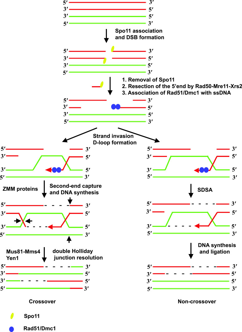
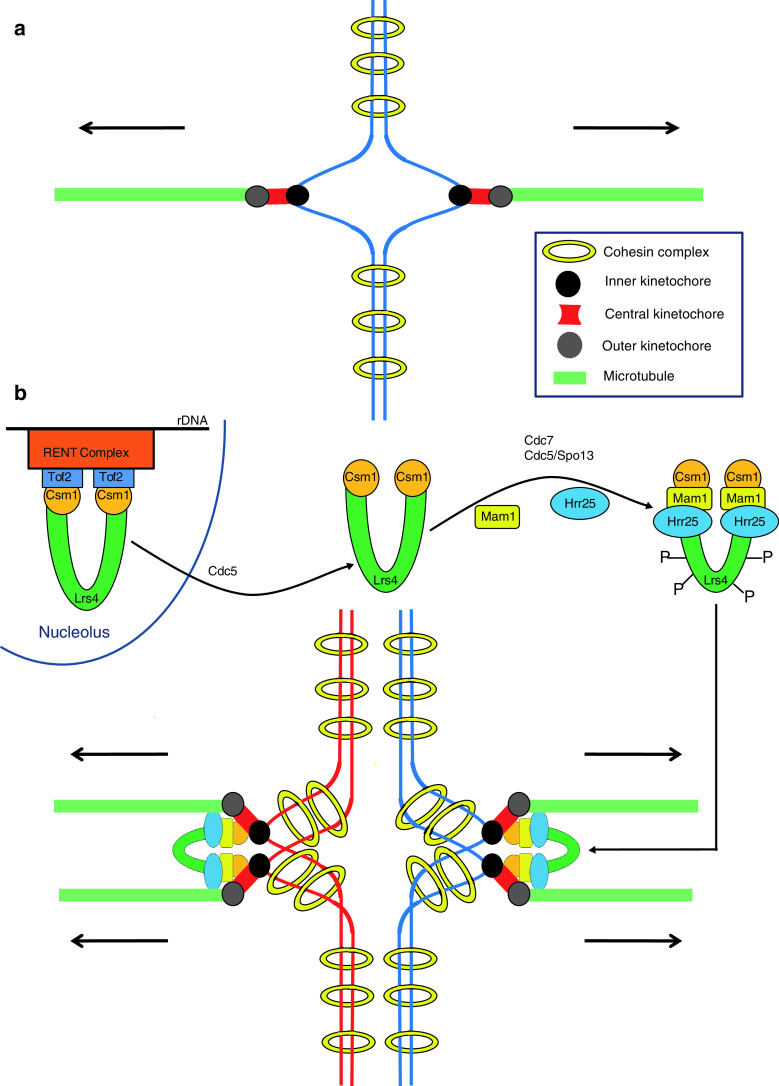
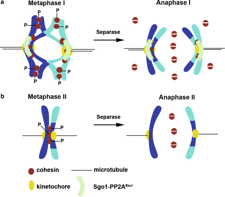
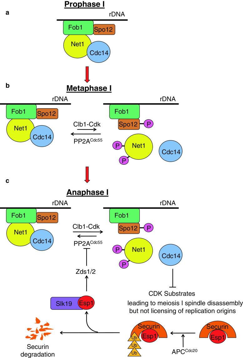
Maintenance of ploidy in sexually reproducing organisms requires a specialized form of cell division called meiosis that generates genetically diverse haploid gametes from diploid germ cells. Meiotic cells halve their ploidy by undergoing two rounds of nuclear division (meiosis I and II) after a single round of DNA replication. Research in Saccharomyces cerevisiae (budding yeast) has shown that four major deviations from the mitotic cell cycle during meiosis are essential for halving ploidy. The deviations are (1) formation of a link between homologous chromosomes by crossover, (2) monopolar attachment of sister kinetochores during meiosis I, (3) protection of centromeric cohesion during meiosis I, and (4) suppression of DNA replication following exit from meiosis I. In this review we present the current understanding of the above four processes in budding yeast and examine the possible conservation of molecular mechanisms from yeast to humans.


Figures

Fig. 1
Fig. 1
Pathway for meiotic recombination in budding yeast. Recombination is initiated by creation of DSBs in DNA by Spo11. The 5′ ends are resected by the Rad50-Mre11-Xrs2 (RMX) complex acting together with Com2/Sae1 and Exo1. Nucleoprotein filaments formed by association of recombinases Rad51 and meiosis-specific Dmc1 with single-stranded DNA scan the genome for homologous sequences. When a match is found the 3′ end base pairs with the complementary strand displacing the other strand to form a D-loop. At this stage there are two possible routes. In the SDSA pathway the invading strand dissociates from the homologous strand and re-anneals with its complementary strand leading to a non-crossover. A crossover is formed when the displaced strand base-pairs with the strand complementary to the invading strand to form a dHJ which is subsequently resolved by a pair of nick and ligation reactions by resolvases Mus81/Mms4 and Yen1
Fig. 2
Fig. 2
Monopolar attachment of sister kinetochores during meiosis I. a Cohesion between sister chromatids (blue) is mediated by cohesin (yellow rings). A specialized structure called the kinetochore is formed at the centromere on each chromatid. The kinetochore is composed of three distinct layers the inner (black), the central (red) and the outer (gray) kinetochores. Sister kinetochores bind to microtubules (green rods) emanating from opposite spindle poles during mitosis (bi-orientation). b Csm1 and Lrs4 proteins localize to the nucleolus via their binding to Tof2 which is bound to rDNA via its association with the RENT (regulator of nucleolar silencing and telophase) complex. After exit from pachytene, Cdc5 triggers release of Csm1 and Lrs4 from the nucleolus. The Csm1/Lrs4 complex associates with meiosis-specific Mam1 and casein kinase-1 (Hrr25) subunits to form the monopolin complex. After nucleolar release, Lrs4 is phosphorylated by Dbf4-dependent kinase Cdc7 and Cdc5 acting together with meiosis-specific protein Spo13. The monopolin complex associates with sister kinetochores and crosslinks them such that they bind to microtubules from the same spindle pole. For the sake of simplicity chiasmata are not shown
Fig. 3
Fig. 3
Protection of centromeric cohesion during meiosis I. a During metaphase I, Sgo1 targets the protein phosphatase PP2ARts1 to the kinetochore which shields centromeric cohesin from phosphorylation by casein kinase-1 (Hrr25) and Dbf4-dependent kinase (DDK). Separase cleaves arm cohesin (phosphorylated) to resolve chiasmata during anaphase I, but does not cleave centromeric cohesin (dephosphorylated). b Loss of Sgo1/PP2ARts1 function after meiosis I results in phosphorylation of centromeric cohesin. Separase gets reactivated during meiosis II and cleaves centromeric cohesin to separate dyad chromosomes
Fig. 4
Fig. 4
The FEAR network regulates exit from meiosis I. a Until prophase I, Cdc14 is bound to the nucleolus via its physical interaction with its competitive inhibitor Net1 which is enhanced by the replication fork barrier protein Fob1. b Increase in Cdk activity following exit from pachytene promotes phosphorylation of Net1, but this is opposed by the protein phosphatase PP2ACdc55 restraining Cdc14 release from the nucleolus. This state persists until metaphase I. c During anaphase I, separase is activated by destruction of its inhibitor securin which is targeted for proteasomal degradation by APCCdc20. Activated separase in association with Slk19 inhibits PP2ACdc55 via Zds1/2 proteins which results in phosphorylation of Net1 and Cdc14 release from the nucleolus. In addition, activation of Spo12 by Cdk-mediated phosphorylation weakens the effect of Fob1 in stabilizing Net1–Cdc14 interaction. The Cdc14 released antagonizes Cdk activity which is sufficient to cause spindle disassembly but not licensing of DNA replication origins

References

    1. Hassold T, Hunt P. To err (meiotically) is human: the genesis of human aneuploidy. Nat Rev Genet. 2001;2:280–291. - PubMed
    1. Gruber S, Haering CH, Nasmyth K. Chromosomal cohesin forms a ring. Cell. 2003;112:765–777. - PubMed
    1. Haering CH, Farcas AM, Arumugam P, Metson J, Nasmyth K. The cohesin ring concatenates sister DNA molecules. Nature. 2008;454:297–301. - PubMed
    1. Haering CH, Lowe J, Hochwagen A, Nasmyth K. Molecular architecture of SMC proteins and the yeast cohesin complex. Mol Cell. 2002;9:773–788. - PubMed
    1. Kassir Y, Adir N, Boger-Nadjar E, Raviv NG, Rubin-Bejerano I, Sagee S, Shenhar G. Transcriptional regulation of meiosis in budding yeast. Int Rev Cytol. 2003;224:111–171. - PubMed

Publication types

Substances

LinkOut - more resources