. 2012 Mar;25(1):103-34.
doi: 10.1293/tox.25.103.

Histopathology of incidental findings in beagles used in toxicity studies

Histopathology of incidental findings in beagles used in toxicity studies

Junko Sato et al. J Toxicol Pathol. 2012 Mar.

Abstract

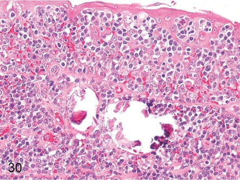
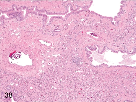
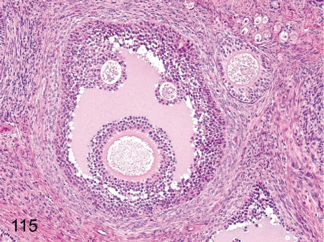
The purpose of our publication is to widely communicate the pictures of spontaneous findings occurring in beagles. Spontaneous arteritis occurs commonly in beagles. Frequent sites of arteritis are the heart, spleen, pancreas, epididymis and spinal cord. Morphological similarities between spontaneous and drug-induced arterial lesions may cause confusion when evaluating vascular toxicity of chemicals such as vasodilating agents. Focal and minimal inflammatory lesions are occasionally seen in the lung and may be associated with aspiration of food particles or of unknown causes. A cystic change with copious mucin production occurs occasionally in the mucosal epithelium of the gall bladder. Nesidioblastosis is seen rarely in the pancreas of beagles. C-cell complex and lymphocytic thyroiditis are common thyroid lesions. Spontaneous focal hypospermatogenesis and lobular Sertoli-cell-only seminiferous tubules occurring frequently in beagles must be distinguished from drug-induced damage of the seminiferous tubules in toxicity studies. The morphological differences of the female genital system in each cycle need to be understood; therefore, we present the normal features of the cyclic changes of the female genital organs. Further, we provide more information on spontaneous findings in beagles for exact diagnoses in toxicity studies.

Keywords: background; beagle; histopathology; incidental; spontaneous.


Figures

Fig. 1.
Fig. 1.
Heart: Blood cyst in the atrioventricular valve. A blood cyst surrounded by endothelia occurs occasionally in the atrioventricular valve, although anatomically, the cardiac valves are nonvascular tissue. The pathogenesis is unknown.
Fig. 2.
Fig. 2.
Heart: Hemorrhage in the atrioventricular valve. Hemorrhage without inflammation or hemosiderosis occurs occasionally in the atrioventricular valve, suggesting that the change is agonal hemorrhage during the euthanasia and exsanguination.
Fig. 3.
Fig. 3.
Heart: Thickening of the intramural arterial wall. Thickening of the wall is due to proliferation of intimal components. The arterial change occurs occasionally in the right auricle.
Fig. 4.
Fig. 4.
Heart: Proliferation of the epicardial mesothelium. Focal papillary proliferation of single layered mesothelial cells occurs occasionally in the right or left atrial epicardium. The change is thought to be a reactive response to physical irritation.
Fig. 5.
Fig. 5.
Heart: Hemorrhage in the endocardium. Slight focal hemorrhage in the endocardium and subendocardium occurs occasionally and is mainly considered an agonal change. It is necessary to distinguish this agonal change from drug-induced hemorrhage by certain drugs such as minoxidil, theobromine and other vasodilating drugs.
Fig. 6.
Fig. 6.
Heart: Focal inflammatory cell infiltration in the myocardium. Focal accumulation of inflammatory cells consisting of mononuclear cells occurs occasionally. The change is sometimes accompanied by slight and focal myocardial necrosis.
Fig. 7.
Fig. 7.
Heart: Fatty infiltration in the myocardium. Adipose tissues are present in the myocardium. This age-related change occurs occasionally in beagles older than one year.
Fig. 8.
Fig. 8.
Artery: Arteritis in the coronary artery. This figure is arteritis of the right coronary artery in the coronary sulcus. The most frequent site of arteritis in beagles is the extramural atrial branch of the right coronary artery. It is difficult to distinguish spontaneously occurring vasculitis from drug-induced vasculitis resulting from such things as minoxidil, theobromine and other vasodilating drugs.
Fig. 9.
Fig. 9.
Artery: Arteritis in the spleen. Arteritis is less frequently seen in the spleen as an isolated lesion or part of polyarteritis. The change is characterized by fibrous thickening with inflammatory cell infiltration into the tunica intima and fibrinoid necrosis of the tunica media.
Fig. 10.
Fig. 10.
Artery: Arteritis in the pancreas. The artery in the pancreas is one of the arteries with a high incidence of arteritis.
Fig. 11.
Fig. 11.
Artery: Arteritis in the spinal cord. Arteritis is seen in the spinal subarachnoid cavity.
Fig. 12.
Fig. 12.
Artery: Arteritis in the epididymis. The epididymal artery is one of the arteries with a high incidence of arteritis, often in the form of periarteritis.
Fig. 13.
Fig. 13.
Aorta: Mineralization in the aortic wall. Mineralization in the tunica media of the aorta is rare and focal in young beagles.
Fig. 14.
Fig. 14.
Lymph node: Blood absorption. Erythrophagia is common in the lymphatic sinus undergoing blood absorption as an agonal change, especially in the mesenteric lymph node.
Fig. 15.
Fig. 15.
Lymph node: Granuloma. A focal lymphohistiocytic response occurs occasionally in the lymph node. The cause is unclear, but it is thought to be a nonspecific reactive change to some irritation such as inflammation, foreign body and parasite. See also Fig. 17.
Fig. 16.
Fig. 16.
Lymph node: Increased number of pigment-laden macrophages. Brown pigment-laden macrophages accumulate predominantly in the medullary region, especially in the mandibular lymph node. Tattoo pigments for animal discrimination occasionally migrate to the mandibular lymph node.
Fig. 17.
Fig. 17.
Lymph node: Foreign body granuloma. Macrophages accumulate and form multinucleated giant cells around foreign bodies such as hair fragments or parasites. Granuloma caused by hair fragments occurs occasionally in intravenous injection studies.
Fig. 18.
Fig. 18.
Lymph node: Increased number of foam cells in the sinuses. The number of foam cells (foamy macrophages) occasionally increases in the lymphatic sinuses, especially in the mesenteric lymph node.
Fig. 19.
Fig. 19.
Thymus: Involution (physiological atrophy). A decrease in lymphoid cells from both the cortex and medulla with replacement by adipose tissues causes reduction in size and organ weight. Thymic involution is common in beagles older than one year old.
Fig. 20.
Fig. 20.
Thymus: Proliferation of the thymic epithelium. The change is characterized by proliferation of prominent tubular structures lined by the cuboidal epithelium. It may be associated with thymic atrophy (involution).
Fig. 21.
Fig. 21.
Thymus: Cyst. Solitary or multilocular cysts are associated with remnants of embryonic ducts connecting the parathyroid and thymus. The cysts are covered by a cuboidal epithelium, partially ciliated, and contain the same eosinophilic proteinic substances as parathyroid cysts (Fig. 131).
Fig. 22.
Fig. 22.
Thymus: Lymph follicle formation in the medulla. A germinal center is commonly formed in the medulla. The change is known to be associated with myasthenia gravis in humans, whereas it is irrelevant and incidental in beagles. Vaccination is considered to be attributable to the change, so attention must be paid to the immunologic failure or overresponse if the follicle formation is severe.
Fig. 23.
Fig. 23.
Spleen: Accessory spleen. The aberration of splenic tissue separated from the original body and surrounding capsule occurs occasionally in the greater omentum and mesenterium as a congenital anomaly.
Fig. 24.
Fig. 24.
Spleen: Enhanced extramedullary hematopoiesis. An increase in hematopoietic cells, predominantly erythrocytic series, occurs occasionally in the red pulp under normal conditions.
Fig. 25.
Fig. 25.
Spleen: Gandy-Gamna body. A focus consisting of relatively dense fibrous tissue or collagenous fibers impregnated with iron pigment and calcium salts is common usually in the splenic hilum and probably results from organization and scarring of the sites where a small perivascular hemorrhage occurred.
Fig. 26.
Fig. 26.
Spleen: Necrosis of the splenic follicle. This is an unusual lesion that shows coagulative necrosis of the germinal center.
Fig. 27.
Fig. 27.
Spleen: Pigment deposition in the red pulp. This is a common lesion that shows an increase in brown pigment-laden macrophages usually containing hemosiderin.
Fig. 28.
Fig. 28.
Spleen: Nodular hyperplasia (lymphoid follicle). Nodular hyperplasia is essentially focal proliferation of various types of splenic components compressing slightly normal splenic tissues, although a large aggregation of lymphocytes is the main feature. The nodule takes an appearance like a giant follicle without the normal structures of a lymph follicle such as a central artery, periarterial lymphoid sheath (PALS) or germinal center. The follicles out of the nodule are normal. There is no evidence to support that this lesion progresses to lymphoma.
Fig. 29.
Fig. 29.
Bone marrow: Lymph follicle formation. Focal accumulation of lymphocytes occurs occasionally in the femur and sternum bone marrow. It generally takes on the appearance of a primary follicle.
Fig. 30.
Fig. 30.
Tonsilla: Mineralization in the lymphoid tissue. Mineralization sometimes occurs in the tonsilla.
Fig. 31.
Fig. 31.
Tonsilla: Neutrophilic migration in the mucosa. Neutrophils frequently migrate into the mucosal epithelium of the tonsilla.
Fig. 32.
Fig. 32.
Larynx: Focal inflammatory cell infiltration. Focal inflammatory cell infiltration occurs occasionally in the vocal fold.
Fig. 33.
Fig. 33.
Larynx: Erosion/ulcer of the mucosa. Erosion and ulcer of the larynx mucosa are rare.
Fig. 34.
Fig. 34.
Trachea: Focal inflammatory cell infiltration. Focal lymphocytic infiltration occurs occasionally in the tracheal mucosa. The mucosal epithelial cells are often intact, as shown in this case.
Fig. 35.
Fig. 35.
Trachea: Hyperplasia of the mucosal epithelium in the tracheal bifurcation. Hyperplasia of the mucosal epithelium is frequently seen in the region near the bifurcation.
Fig. 36.
Fig. 36.
Trachea: Squamous metaplasia in the tracheal bifurcation. Squamous metaplasia of the mucosal epithelium leading from a hyperplastic lesion occurs occasionally in the region near the bifurcation. The tracheal bifurcation is predisposed to hyperplasia (Fig. 35) and squamous metaplasia because this region is most likely to receive the physiological irritant effects.
Fig. 37.
Fig. 37.
Lung: Accumulation of foam cells in the alveolus. Focal accumulation of foam cells without other inflammatory cells is frequently seen in the alveoli.
Fig. 38.
Fig. 38.
Lung: Lobar hypoplasia. Lobar atelectasis (stenosis of the bronchiole and alveoli) and bronchial cartilage aplasia occur occasionally. Grossly, the affected lobe is very small, suggesting congenital hypoplasia.
Fig. 39.
Fig. 39.
Lung: Focal inflammatory cell infiltration. Focal lymphocytic infiltration is common in the terminal bronchiole.
Fig. 40.
Fig. 40.
Lung: Focal hemorrhage in the alveolus. Focal hemorrhage occurs occasionally in the alveoli. Hemorrhage without inflammatory cell infiltration is suggestive of an agonal change.
Fig. 41.
Fig. 41.
Lung: Inflammatory cell infiltration in the alveolus. Inflammatory cell infiltration with/without hemorrhage occurs occasionally.
Fig. 42.
Fig. 42.
Lung: Fibrosis of the alveolar wall. Focal fibrous thickening of the alveolar wall is common. The change is mainly localized around the bronchiole of the right anterior lobe.
Fig. 43.
Fig. 43.
Lung: Osseous metaplasia. Osseous metaplasia presumably caused by differentiation from fibroblasts to osteoblasts occurs occasionally and is similar to that seen in rats and mice. The frequency of this change in dogs is lower than that of rats.
Fig. 44.
Fig. 44.
Lung: Squamous metaplasia of the bronchial epithelium. Focal squamous metaplasia of the bronchial epithelium with/without inflammation occurs occasionally. The change is accompanied by focal epithelial hyperplasia.
Fig. 45.
Fig. 45.
Lung: Thrombus. Thrombosis is frequently seen in the pulmonary vessels of beagles used in toxicity studies by intravenous injection.
Fig. 46.
Fig. 46.
Lung: Foreign body granuloma. Foreign bodies are surrounded by macrophages and lymphocytes. This change results from aspiration of stomach contents or food particles (arrow).
Fig. 47.
Fig. 47.
Lung: Organized thrombus and foreign body. Stenosis of the vascular lumen by a fibrous organization of thrombus containing hair fragments is rare. The hair fragment may enter during intravenous injection.
Fig. 48.
Fig. 48.
Tongue: Foreign body granuloma. Foreign body granuloma caused by a hair fragment stuck on the tongue occurs occasionally in the lamina propria or muscle layer.
Fig. 49.
Fig. 49.
Tongue: Focal infiltration of macrophages in the muscle layer. Focal infiltration of macrophages occurs occasionally in the muscle layer.
Fig. 50.
Fig. 50.
Esophagus: Focal atrophy of the esophageal gland. Focal atrophy of the esophageal gland occurs occasionally.
Fig. 51.
Fig. 51.
Esophagus: Hypertrophy of the esophageal gland. Hypertrophic acinar cells including abundant mucus are rare in the esophageal gland.
Fig. 52.
Fig. 52.
Esophagus: Focal inflammatory cell infiltration in the lamina propria. Focal lymphocytic infiltration is common in the lamina propria.
Fig. 53.
Fig. 53.
Stomach: Infection by Helicobacter heilmannii in the gastric mucosa. Helicobacter spp. infection occurs occasionally in the lumen of the pyloric gland without inflammation. Many beagles have Helicobacter heilmannii in their stomach.
Fig. 54.
Fig. 54.
Stomach: Mineralization in the lamina propria. Focal mineralization is common in the lamina propria.
Fig. 55.
Fig. 55.
Duodenum: Hyperplasia of the lymphoid tissue. Hyperplasia of the lymphoid tissue occurs occasionally in not only the duodenum but other intestinal tracts. Development of the intestinal lymphoid tissue varies among individual animals.
Fig. 56.
Fig. 56.
Duodenum: Dilatation of the crypt. Dilatation of the crypt filled with/without cell debris or inflammatory cell infiltration is frequently seen. The change most frequently occurs in the duodenum but also occurs in the other intestines.
Fig. 57.
Fig. 57.
Duodenum: Ectopic pancreatic tissue. Ectopic pancreatic tissue is frequently seen in the submucosa near the major duodenal papilla.
Fig. 58.
Fig. 58.
Jejunum: Ectopic fundic gland. An ectopic fundic gland is rarely seen in the jejunum. The ectopic fundic gland is composed of normal fundic tissue.
Fig. 59.
Fig. 59.
Cecum: Granular cell tumor. Large polygonal eosinophilic cells infiltrate into the lamina propria and submucosa of the cecum. The tumor cells have fine granular cytoplasm. This tumor is rare in beagles.
Fig. 60.
Fig. 60.
Salivary gland: Focal inflammatory cell infiltration. Focal lymphocytic infiltration, occasionally accompanied by lymph follicle formation, is common around the ducts or acini.
Fig. 61.
Fig. 61.
Salivary gland: Focal fibrosis. Focal fibrosis with atrophy and loss of the acini is frequently seen, especially in the parotid gland.
Fig. 62.
Fig. 62.
Salivary gland: Salivary calculus. A small mineralized calculus occurs occasionally in a duct of a relatively large caliber.
Fig. 63.
Fig. 63.
Salivary gland: Increase in the mucous gland. The mucous epithelium increases occasionally in the parotid gland, though the parotid gland of the beagles is composed mainly of a serous epithelium.
Fig. 64.
Fig. 64.
Liver: Accumulation of glycogen in hepatocytes. The change is common in beagles and characterized by a clear appearance of hepatic cytoplasm after formalin fixation. The quantity of glycogen in cytoplasm varies among individuals under normal conditions.
Fig. 65.
Fig. 65.
Liver: Microgranuloma. Microgranuloma is accumulation of inflammatory cells, mainly macrophages, lymphocytes and a small number of neutrophils, and may be associated with minute necrosis of hepatocytes. It is found frequently.
Fig. 66.
Fig. 66.
Liver: Pigment deposition in hepatocytes. Lipofuscin pigments are common in centrilobular hepatocytes of young beagles but are particularly prominent in older dogs.
Fig. 67.
Fig. 67.
Liver: Focal necrosis of hepatocytes. A necrotic focus of hepatocytes with inflammatory cell infiltration occurs occasionally without any apparent cause.
Fig. 68.
Fig. 68.
Liver: Pigment deposition in Kupffer cells. Kupffer cells occasionally engulf brown pigments in the sinusoid. The pigments are considered to be hemosiderin.
Fig. 69.
Fig. 69.
Liver: Increased number of Ito cells with lipid accumulation. Some Ito cells containing lipid droplets are sporadically present along the sinusoid.
Fig. 70.
Fig. 70.
Gall bladder: Cystic mucinous hyperplasia of the mucosal epithelium. A cystic change or cystic hyperplasia of the mucosal epithelium with copious mucin production occurs occasionally.
Fig. 71.
Fig. 71.
Gall bladder: Hyperplasia of the mucosal epithelium. Hyperplasia of the mucosal epithelium with lymphocytic infiltration in the lamina propria is rare. Unlike Fig. 70, neither copious mucin production nor cystic change is found.
Fig. 72.
Fig. 72.
Gall bladder: Hyperplasia of the lymphoid tissue. Prominent lymphoid tissues with/without lymph follicle formation are common in the lamina propria.
Fig. 73.
Fig. 73.
Pancreas: Focal fibrosis. Focal fibrosis with acinar atrophy and/or loss occurs occasionally.
Fig. 74.
Fig. 74.
Pancreas: Apoptosis of acinar cells. Apoptosis of acinar cells occurs occasionally in the pancreas in beagles. This case has diffuse and relatively abundant apoptosis in the acini.
Fig. 75.
Fig. 75.
Pancreas: Nesidioblastosis. Ductular cells irregularly proliferate between acinar cells. Irregularly shaped islets and small clusters of endocrine cells closely associate with ductules or intercalated ductules. The change is rare and appears as a proliferative lesion of the pancreas.
Fig. 76.
Fig. 76.
Kidney: Glomerular lipidosis. Focal and segmental lipidosis occurs occasionally in the glomeruli. Mesangial cells contain lipid droplets, and are frequently accompanied by hyaline droplets.
Fig. 77.
Fig. 77.
Kidney: Osseous metaplasia. Osseous metaplasia rarely occurs in the glomerulus and interstitium.
Fig. 78.
Fig. 78.
Kidney: Immature glomerulus. Capillary formation is indistinct, whereas epithelial cells are prominent in an immature glomerulus. Immature glomerulus is more frequently seen in the outer layer of the cortex, reflecting delayed maturation of the glomeruli in the outer cortex than those in the deeper layer.
Fig. 79.
Fig. 79.
Kidney: Focal segmental sclerosis of the glomerulus. Segmental eosinophilic nodule containing some mesangial cells and matrix in one or a few glomeruli is rare. A lesion such as that resulting from collapse of the capillary loops due to the proliferation of mesangial cells and increase in matrix is called sclerosis.
Fig. 80.
Fig. 80.
Kidney: Renal dysplasia. A typical lesion of renal dysplasia consists of a scarring focus containing mesenchymal cells, immature renal tubules (large arrows), small-sized glomeruli (small arrows) and dilatation of the collecting tubule (arrowheads) in the medulla to the cortex. This change is rare.
Fig. 81.
Fig. 81.
Kidney: Regeneration of the tubular epithelium. Focal regenerative changes of damaged tubules are detected frequently as basophilic tubules with a high nuclear density in the cortex or outer stripe of the renal medulla. Inflammatory cell infiltration occurs occasionally around the basophilic tubules.
Fig. 82.
Fig. 82.
Kidney: Focal inflammatory cell infiltration in the interstitium. Focal lymphocytic infiltration is common in the interstitium. The infiltrated cells often show a plasmacytic morphology with basophilic cytoplasm.
Fig. 83.
Fig. 83.
Kidney: Hyaline cast. A solitary cast occurs occasionally in the distal segment or collecting tubule in the outer medulla, but it is not associated with chronic progressive nephropathy as in rats.
Fig. 84.
Fig. 84.
Kidney: Focal interstitial fibrosis. Focal interstitial fibrosis occurs occasionally and is accompanied by interstitial inflammation, pyelitis and infarction. The fibrous tissue may occasionally contain some atrophic tubules and hyaline casts.
Fig. 85.
Fig. 85.
Kidney: Brown pigment deposition in the tubular epithelium. Brown pigment deposition occurs occasionally in the proximal tubular epithelium. The deposited pigments are considered to be lipofuscin.
Fig. 86.
Fig. 86.
Kidney: Fatty change of the proximal tubular epithelium. Tubules containing fine lipid droplets are frequently seen in the epithelia of the straight portion of the proximal tubules in most female dogs.
Fig. 87.
Fig. 87.
Kidney: Inclusion bodies in the collecting tubular epithelium. Basophilic/eosinophilic cytoplasmic inclusion bodies occur occasionally in the renal papillary collecting tubules. These inclusion bodies are frequently accompanied by pyelitis at the same time.
Fig. 88.
Fig. 88.
Kidney: Mineralization in the medulla. Small mineralized calculus is frequently seen in the renal papillary collecting tubules.
Fig. 89.
Fig. 89.
Kidney: Focal hyperplasia of the pelvic epithelium. Focal hyperplasia of the transitional epithelium (urothelium) covering the papilla with inflammation occurs occasionally, but the lesion without inflammation, as in this case, is rare.
Fig. 90.
Fig. 90.
Kidney: Pyelitis. Focal lymphocytic infiltration including lymph follicle formation is frequently seen in the lamina propria of the renal pelvis. Pyelitis may progress to pyelonephritis (Figs. 91, 92) by ascending spread of inflammation.
Fig. 91.
Fig. 91.
Kidney: Pyelonephritis. A severe case of pelvic inflammation penetrating all mucosal regions and the papillary interstitium with tubular damage occurs occasionally.
Fig. 92.
Fig. 92.
Kidney: Interstitial nephritis. This tubulointerstitial inflammation can be assumed to be accompanied by pyelitis because the lesion distributed along nephron is suggested to be an ascending spread of inflammatory lesions from the pelvis.
Fig. 93.
Fig. 93.
Urinary bladder: Round ligament of the urinary bladder. A fibromuscular band with mineralization is rarely attached to the urinary bladder. This is a remnant of the umbilical artery.
Fig. 94.
Fig. 94.
Testis: Immature. Immature seminiferous tubules occur occasionally in young beagles aged 6-7 months. Only a small number of spermatids are present, whereas development up to the spermatocytes completes in the seminiferous tubules.
Fig. 95.
Fig. 95.
Testis: Hypospermatogenesis. This is a common spontaneous change characterized by scarce spermatocytes and spermatids. It is a focal failure of spermatogenesis in the seminiferous tubules.
Fig. 96.
Fig. 96.
Testis: Sertoli-cell-only tubule. A segmental lesion of Sertoli-cell-only-tubules is common. The change is probably segmental hypoplasia of the seminiferous tubules during testicular development. When seminiferous tubules sustain a permanent damage, the end-stage feature shows Sertoli-cell-only tubules.
Fig. 97.
Fig. 97.
Testis: Appearance of multinucleated giant cells. Focal or sporadic multinucleated giant cell formation occurs occasionally in the seminiferous tubules.
Fig. 98.
Fig. 98.
Testis: Vacuolation of Sertoli cells. Sporadic vacuolation of Sertoli cells is frequently seen in the seminiferous tubules.
Fig. 99.
Fig. 99.
Testis: Spermatocele. Aggregation of spermatozoa occurs occasionally in some parts of the seminiferous tubules. This change is caused by disturbance of spermatozoa flow due to seminiferous tubular damage.
Fig. 100.
Fig. 100.
Testis: Retention of sperm in the seminiferous tubules. Retention of spermatids is frequently associated with tubular damage.
Fig. 101.
Fig. 101.
Epididymis: Immature. The ductal epithelia are small and flat, and lumens are narrow. The interstitium also consists of immature fibrous tissue with high nuclear density.
Fig. 102.
Fig. 102.
Epididymis: Cell debris in the epididymal lumen. Cell debris of spermatocytes or spermatids occurs occasionally in the epididymal lumen in normal beagles. It is prominent when the testis has some seminiferous tubular damage.
Fig. 103.
Fig. 103.
Epididymis: Intraepithelial lumen. An intraepithelial lumen is frequently seen and is lined by ciliated cells.
Fig. 104.
Fig. 104.
Epididymis: Mineralization of the epithelium. Mineral deposition occurs occasionally in the epididymal epithelia.
Fig. 105.
Fig. 105.
Epididymis: Spermatocele. A mass of accumulated spermatozoa occurs occasionally in the lumen. Granulomatous inflammation (spermatic granuloma) can occur when the basal lamina ruptures.
Fig. 106.
Fig. 106.
Epididymis: Spermatic granuloma (sperm granuloma). Foreign body granuloma occurs occasionally in the interstitium and contains a mass of spermatozoa surrounded by macrophages and lymphocytes.
Fig. 107.
Fig. 107.
Epididymis: Focal inflammatory cell infiltration in the interstitium. Focal lymphocytic infiltration is common in the interstitium.
Fig. 108.
Fig. 108.
Prostate: Immature. The glandular epithelia are small and flat, and lumens are narrow because of no prostatic secretion. Maturation of the prostatic gland varies in young beagles aged 6-7 months.
Fig. 109.
Fig. 109.
Prostate: Focal inflammatory cell infiltration. Focal lymphocytic infiltration is frequently seen in the interstitium.
Fig. 110.
Fig. 110.
Female reproductive system: Proestrus. Ovary (containing antral follicle), uterus, vagina, mammary gland.
Fig. 111.
Fig. 111.
Female reproductive system: Estrus. Ovary (containing postovulatory corpus luteum), uterus, vagina, mammary gland
Fig. 112.
Fig. 112.
Female reproductive system: Metestrus. Ovary (containing functional corpus luteum), uterus, vagina, mammary gland
Fig. 113.
Fig. 113.
Female reproductive system: Metestrus to diestrus. Ovary (containing involuting corpus luteum), uterus, vagina, mammary gland
Fig. 114.
Fig. 114.
Female reproductive system: Diestrus. Ovary (containing previously formed corpus luteum), uterus, vagina, mammary gland
Fig. 115.
Fig. 115.
Ovary: Polyovular follicle. Multiple ova occur occasionally in one ovarian follicle. Each oocyte has its own pellucid zone and radiate corona.
Fig. 116.
Fig. 116.
Ovary: Mineralization. Primordial follicles and early secondary follicles occur occasionally mineralized. Mineralization presumably occurs in the necrotized oocytes accompanied by follicular atresia.
Fig. 117.
Fig. 117.
Ovary: Hyperplasia of the rete ovarii. The embryonic remnants of rete ovarii are frequently seen in the hilum of the ovary and occasionally undergo glandular hyperplasia.
Fig. 118.
Fig. 118.
Ovary: Cyst. A large cyst occurs occasionally in the ovarian parenchyma. The luminal surface is lined by a single layer of flat or cuboidal ciliated epithelial cells. This cyst is probably a paroophoritic cyst.
Fig. 119.
Fig. 119.
Ovary: Corpus luteum with cyst. This change occurs occasionally in the corpus luteum. The cause of this change is probably a luteinized unruptured follicle.
Fig. 120.
Fig. 120.
Uterus: Placenta-like endometrial hyperplasia. Cystic epithelial proliferation of the endometrium separated into two layers is formed in the uterus in beagles. This finding occurs occasionally and is suggestive of pseudopregnancy.
Fig. 121.
Fig. 121.
Pituitary: Cyst. Pituitary cysts are frequently seen in each lobe (anterior, intermediate, posterior) but are most often found in the anterior lobe.
Fig. 122.
Fig. 122.
Pituitary: Focal inflammatory cell infiltration in the posterior lobe. Focal lymphocytic infiltration occurs occasionally around the vessel or other parenchyma.
Fig. 123.
Fig. 123.
Thyroid (Parathyroid): Ectopic thymic tissue. Thymic tissue is frequently seen in the thyroid or parathyroid tissue. The parathyroid, thyroid and thymic primordia derive from the same primordium, the pharyngeal pouches, during embryonic development.
Fig. 124.
Fig. 124.
Thyroid: C-cell complex. Solid islands of C-cell-like clear cells are frequently seen in the thyroid tissue. They are considered to be remnants of ultimobranchial bodies formed before differentiation into follicular cells and C-cells.
Fig. 125.
Fig. 125.
Thyroid: Cyst. Large cysts occur occasionally in the thyroid parenchyma. The cysts are lined by a cuboidal epithelium that is often ciliated like a parathyroid cyst (Fig. 131).
Fig. 126.
Fig. 126.
Thyroid (parathyroid): Ultimobranchial remnant. The duct is a remnant of the ultimobranchial body in the thyroid or parathyroid gland. It is lined by a few layered squamous epithelial cells and often cystic. The incidence in beagles is lower than that in rats.
Fig. 127.
Fig. 127.
Thyroid: Focal atrophy of follicles. This change is rare. The follicles are focally small in size and contain a small amount of colloid inside with abundant connective tissue between each atrophic follicle.
Fig. 128.
Fig. 128.
Thyroid: Giant follicle. Enlarged follicles aggregate and mildly compress the peripheral parenchyma. Papillary proliferation of the epithelial cells and an increase in small follicles are absent; these may be the differential points from focal follicular hyperplasia. Giant follicles are rare.
Fig. 129.
Fig. 129.
Thyroid: Mineralization of follicles. Mineralization occurs occasionally in the follicle.
Fig. 130.
Fig. 130.
Thyroid: Lymphocytic thyroiditis. This change is frequently seen and is characterized by lymphocytic infiltration often forming a prominent germinal center in the interstitium. The follicles are destroyed and infiltrated also by lymphocytes, and residual follicles are lined with hypertrophic epithelial cells. The etiology of this lesion is considered to be an autoimmune disorder.
Fig. 131.
Fig. 131.
Parathyroid: Cyst (Kürsteiner’s cyst). The cyst is lined by a cuboidal epithelium, often ciliated, and contains proteinous substance in the lumens. This cyst is an embryonal remnant of the duct connecting the parathyroid-thymus tissue in the III and IV pharyngeal pouches.
Fig. 132.
Fig. 132.
Parathyroid: Ectopic cartilaginous tissue. Well differentiated cartilage tissue is rarely formed in the parenchyma.
Fig. 133.
Fig. 133.
Parathyroid: Fatty infiltration. Adipose cells infiltrate sporadically in the interstitium.
Fig. 134.
Fig. 134.
Parathyroid: Fibrosis. Fibrous tissue occasionally increases in the interstitium.
Fig. 135.
Fig. 135.
Adrenal: Accessory adrenocortical tissue. The aberration of adrenocortical tissue separated from the original body and surrounding capsule is called accessory adrenocortical tissue, and it is common inside and outside of the adrenal capsule.
Fig. 136.
Fig. 136.
Adrenal: Vacuolation of the cells of the zona glomerulosa. Accumulation of large lipid droplets containing cells is frequently seen in the zona glomerulosa in beagles.
Fig. 137.
Fig. 137.
Adrenal: Osseous metaplasia. Osseous metaplasia in the adrenal gland is rare. The metaplastic bone is usually beneath the capsule.
Fig. 138.
Fig. 138.
Adrenal: Vacuolation of the cells of the zona fasciculata. Various degrees of zonal vacuolation in the cells of the zona fasciculata occur occasionally, especially near the zona reticularis.
Fig. 139.
Fig. 139.
Adrenal: Extramedullary hematopoiesis. Erythrocytic or granulocytic extramedullary hematopoiesis occurs occasionally in the adrenal sinusoids.
Fig. 140.
Fig. 140.
Adrenal: Pigment deposition in the corticomedullary junction. Deposition of yellow-brown pigments occurs occasionally in the cytoplasm of cortical cells near the corticomedullary junction. The pigments are considered to be ceroid or lipofuscin.
Fig. 141.
Fig. 141.
Brain: Focal inflammatory cell infiltration in the choroid plexus. Lymphoplasmacytes occasionally infiltrate into the interstitium of the choroid plexus.
Fig. 142.
Fig. 142.
Brain: Extramedullary hematopoiesis in the choroid plexus. Hematopoietic cells of granulocytic series with rare megakaryocytes (arrow) occasionally aggregate in the choroid plexus.
Fig. 143.
Fig. 143.
Brain: Mineralization of the meninx. Focal mineralization occurs occasionally in the meninges.
Fig. 144.
Fig. 144.
Brain: Fibrous thickening of the meninx. Fibrous thickening of the meninx may have resulted from chronic inflammation or circulatory disturbance and is very rare.
Fig. 145.
Fig. 145.
Brain: Hamartoma in the callosal sulcus. This rare lesion consists of choroid plexus components that are epithelia, fatty tissues, abundant collagen fibers and small vessels. It is considered that the lesion is possibly an ectopic tissue.
Fig. 146.
Fig. 146.
Spinal cord: Mineralization. Mineralization of the vessel wall in the spinal nerve roots occurs occasionally. This change occurs easily with advancing age.
Fig. 147.
Fig. 147.
Sciatic nerve: Renaut body. An irregular fine fibrous structure in the nerve fiber bundles is common. Its function is thought to be protection of the peripheral nerve fibers from pressure damage.
Fig. 148.
Fig. 148.
Skin: Dermoid cyst. A cyst covered by epidermis with a skin appendage structure occurs occasionally in the dermis. The cyst includes hair sheaths or keratin.
Fig. 149.
Fig. 149.
Skin: Folliculitis. Inflammatory cells infiltrate into the follicle and perifollicular interstitial area. This change is frequently seen in a variety of grades.
Fig. 150.
Fig. 150.
Skin: Parasitic folliculitis. Inflammatory cells infiltrate around follicles affected by parasites. Parasites considered to be Demodex folliculorum exist in the affected follicles.
Fig. 151.
Fig. 151.
Skin: Ulcer. Ulcer is rare as a secondary change associated with folliculitis or other skin lesions.
Fig. 152.
Fig. 152.
Eye: Disarrangement of the retinal structures. A linear folding of the retinal tissue occurs occasionally. The causes of this lesion may be congenital failures of optic fissure closure (retinal coloboma), dysplasia of retinal structures or focal damage of the retina in the fetal developmental stage.
Fig. 153.
Fig. 153.
Eye: Focal inflammatory cell infiltration in the conjunctiva. Lymphocytic infiltration is frequently seen in the subepithelium of the conjunctiva.
Fig. 154.
Fig. 154.
Lacrimal gland: Focal fibrosis. Focal fibrosis of the interstitium with lymphocytic infiltration and acinar atrophy occur occasionally in the lacrimal gland.
Fig. 155.
Fig. 155.
Lacrimal gland: Focal inflammatory cell infiltration. Focal lymphocytic infiltration is common in the interstitium.

References

    1. Jones TC, Hunt RD, King NW. Veterinary Pathology. 6th ed. Lippincott Williams & Wilkins, Baltimore. 1997
    1. Maxie MG. Jubb, Kennedy, and Palmer’s Pathology of Domestic Animals. 5th ed. Elsevier, Philadelphia. 2007
    1. Mohr U, Carlton WW, Dungworth DL, Benjamin SA, Capen CC, Hahn FF. Pathobiology of the Aging Dog. Iowa State University Press, Ames. 2001
    1. Japanese Society of Veterinary Pathology. Colour Atlas of Animal Pathology. Bun-eido Publishing, Tokyo. 2007. (in Japanese).
    1. Gross TL, Ihrke PJ, Walder EJ, Affolter VK. Skin Diseases of the Dog and Cat: Clinical and Histopathologic Dignosis. Blackwell Science, Oxford. 2005