

Is cohesin required for spindle-pole-body/centrosome cohesion?
- PMID: 22482005
- PMCID: PMC3291308
- DOI: 10.4161/cib.18557
Is cohesin required for spindle-pole-body/centrosome cohesion?
Abstract
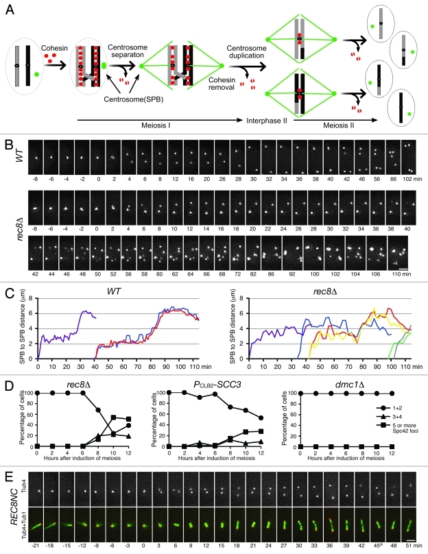
Centrosomes are microtubule-organizing centers that nucleate spindle microtubules during cell division. In budding yeast, the centrosome, often referred to as the spindle pole body, shares structural components with the centriole, the central core of the animal centrosome. The parental centrosome is duplicated when DNA replication takes place. Like sister chromatids tethered together by cohesin, duplicated centrosomes are linked and then separate to form the bipolar spindle necessary for chromosome segregation. Recent studies have shown that cohesin is also localized to the animal centrosome and is perhaps directly involved in engaging paired centrioles. Here we discuss the potential role of cohesin in mediating spindle-pole-body cohesion in the context of yeast meiosis. We propose that the coordination of chromosome segregation with centrosome cohesion and duplication is mediated by the antagonistic interaction between the Aurora kinase and the Polo kinase and that the role of cohesin in centrosome regulation appears to be indirect in budding yeast.
Keywords: SPB cohesion; centrosome; cohesin; meiosis; spindle pole body (SPB).
Figures
Similar articles
-
The Aurora kinase Ipl1 is necessary for spindle pole body cohesion during budding yeast meiosis.J Cell Sci. 2011 Sep 1;124(Pt 17):2891-6. doi: 10.1242/jcs.086652. J Cell Sci. 2011. PMID: 21878496 Free PMC article.
-
The half-bridge component Kar1 promotes centrosome separation and duplication during budding yeast meiosis.Mol Biol Cell. 2018 Aug 1;29(15):1798-1810. doi: 10.1091/mbc.E18-03-0163. Epub 2018 May 30. Mol Biol Cell. 2018. PMID: 29847244 Free PMC article.
-
Centrosome Remodelling in Evolution.Cells. 2018 Jul 6;7(7):71. doi: 10.3390/cells7070071. Cells. 2018. PMID: 29986477 Free PMC article. Review.
-
Centrosomes in mitotic spindle assembly and orientation.Curr Opin Struct Biol. 2021 Feb;66:193-198. doi: 10.1016/j.sbi.2020.11.003. Epub 2020 Dec 6. Curr Opin Struct Biol. 2021. PMID: 33296732 Review.
-
Rad21 is required for centrosome integrity in human cells independently of its role in chromosome cohesion.Cell Cycle. 2010 May;9(9):1774-80. doi: 10.4161/cc.9.9.11524. Epub 2010 May 15. Cell Cycle. 2010. PMID: 20404533
References
LinkOut - more resources
Full Text Sources
Molecular Biology Databases