Linking differences in membrane tension with the requirement for a contractile actomyosin scaffold during exocytosis in salivary glands
- PMID: 22482019
- PMCID: PMC3291323
- DOI: 10.4161/cib.18258
Linking differences in membrane tension with the requirement for a contractile actomyosin scaffold during exocytosis in salivary glands
Abstract
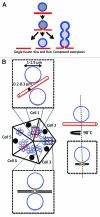
In all the major secretory organs regulated exocytosis is a fundamental process that is used for releasing molecules in the extracellular space. Molecules destined for secretion are packaged into secretory vesicles that fuse with the plasma membrane upon the appropriate stimulus. In exocrine glands, large secretory vesicles fuse with specialized domains of the plasma membrane, which form ductal structures that are in direct continuity with the external environment and exhibit various architectures and diameters. In a recent study, we used intravital microscopy to analyze in detail the dynamics of exocytic events in the salivary glands of live rodents under conditions that cannot be reproduced in in vitro or ex vivo model systems. We found that after the opening of the fusion pore large secretory vesicles gradually collapse with their limiting membranes being completely absorbed into the apical plasma membrane canaliculi within 40-60 sec. Moreover, we observed that this controlled collapse requires the contractile activity of actin and its motor myosin II, which are recruited onto the large secretory vesicles immediately after their fusion with the plasma membrane. Here we suggest that the actomyosin complex may be required to facilitate exocytosis in those systems, such as the salivary glands, in which the full collapse of the vesicles is not energetically favorable due to a difference in membrane tension between the large secretory vesicles and the canaliculi.
Keywords: Secretion; actin; cytoskeleton; exocrine glands; exocytosis; intravital microscopy; membrane tension; myosin; salivary glands.
Figures


Comment on
- Masedunskas A, Sramkova M, Parente L, Sales KU, Amornphimoltham P, Bugge TH, Weigert R. Role for the actomyosin complex in regulated exocytosis revealed by intravital microscopy. Proc Natl Acad Sci USA. 2011;108 doi: 10.1073/pnas.1016778108.
Similar articles
-
The Actomyosin Cytoskeleton Drives Micron-Scale Membrane Remodeling In Vivo Via the Generation of Mechanical Forces to Balance Membrane Tension Gradients.Bioessays. 2018 Sep;40(9):e1800032. doi: 10.1002/bies.201800032. Epub 2018 Aug 6. Bioessays. 2018. PMID: 30080263 Free PMC article. Review.
-
Homeostasis of the apical plasma membrane during regulated exocytosis in the salivary glands of live rodents.Bioarchitecture. 2011 Sep 1;1(5):225-229. doi: 10.4161/bioa.18405. Bioarchitecture. 2011. PMID: 22754613 Free PMC article.
-
Role for the actomyosin complex in regulated exocytosis revealed by intravital microscopy.Proc Natl Acad Sci U S A. 2011 Aug 16;108(33):13552-7. doi: 10.1073/pnas.1016778108. Epub 2011 Aug 1. Proc Natl Acad Sci U S A. 2011. PMID: 21808006 Free PMC article.
-
Exocytosis by vesicle crumpling maintains apical membrane homeostasis during exocrine secretion.Dev Cell. 2021 Jun 7;56(11):1603-1616.e6. doi: 10.1016/j.devcel.2021.05.004. Dev Cell. 2021. PMID: 34102104 Free PMC article.
-
Multiple roles for the actin cytoskeleton during regulated exocytosis.Cell Mol Life Sci. 2013 Jun;70(12):2099-121. doi: 10.1007/s00018-012-1156-5. Epub 2012 Sep 18. Cell Mol Life Sci. 2013. PMID: 22986507 Free PMC article. Review.
Cited by
-
Imaging membrane remodeling during regulated exocytosis in live mice.Exp Cell Res. 2015 Oct 1;337(2):219-25. doi: 10.1016/j.yexcr.2015.06.018. Epub 2015 Jul 6. Exp Cell Res. 2015. PMID: 26160452 Free PMC article. Review.
-
Orchestrated content release from Drosophila glue-protein vesicles by a contractile actomyosin network.Nat Cell Biol. 2016 Feb;18(2):181-90. doi: 10.1038/ncb3288. Epub 2015 Dec 7. Nat Cell Biol. 2016. PMID: 26641716
-
The Actomyosin Cytoskeleton Drives Micron-Scale Membrane Remodeling In Vivo Via the Generation of Mechanical Forces to Balance Membrane Tension Gradients.Bioessays. 2018 Sep;40(9):e1800032. doi: 10.1002/bies.201800032. Epub 2018 Aug 6. Bioessays. 2018. PMID: 30080263 Free PMC article. Review.
-
Homeostasis of the apical plasma membrane during regulated exocytosis in the salivary glands of live rodents.Bioarchitecture. 2011 Sep 1;1(5):225-229. doi: 10.4161/bioa.18405. Bioarchitecture. 2011. PMID: 22754613 Free PMC article.
-
T-Cell Receptor Sequences Identify Combined Coxsackievirus-Streptococci Infections as Triggers for Autoimmune Myocarditis and Coxsackievirus-Clostridia Infections for Type 1 Diabetes.Int J Mol Sci. 2024 Feb 1;25(3):1797. doi: 10.3390/ijms25031797. Int J Mol Sci. 2024. PMID: 38339075 Free PMC article.
References
-
- Burgoyne RD, Morgan A. Secretory granule exocytosis. Physiol Rev. 2003;83:581–632. - PubMed
LinkOut - more resources
Full Text Sources
