Review
. 2012:107:355-415.
doi: 10.1016/B978-0-12-385883-2.00005-9.

Biology of mitochondria in neurodegenerative diseases

Affiliations
Review

Biology of mitochondria in neurodegenerative diseases

Lee J Martin. Prog Mol Biol Transl Sci. 2012.

Abstract

Alzheimer's disease (AD), Parkinson's disease (PD), and amyotrophic lateral sclerosis (ALS) are the most common human adult-onset neurodegenerative diseases. They are characterized by prominent age-related neurodegeneration in selectively vulnerable neural systems. Some forms of AD, PD, and ALS are inherited, and genes causing these diseases have been identified. Nevertheless, the mechanisms of the neuronal degeneration in these familial diseases, and in the more common idiopathic (sporadic) diseases, are unresolved. Genetic, biochemical, and morphological analyses of human AD, PD, and ALS, as well as their cell and animal models, reveal that mitochondria could have roles in this neurodegeneration. The varied functions and properties of mitochondria might render subsets of selectively vulnerable neurons intrinsically susceptible to cellular aging and stress and the overlying genetic variations. In AD, alterations in enzymes involved in oxidative phosphorylation, oxidative damage, and mitochondrial binding of Aβ and amyloid precursor protein have been reported. In PD, mutations in mitochondrial proteins have been identified and mitochondrial DNA mutations have been found in neurons in the substantia nigra. In ALS, changes occur in mitochondrial respiratory chain enzymes and mitochondrial programmed cell death proteins. Transgenic mouse models of human neurodegenerative disease are beginning to reveal possible principles governing the biology of selective neuronal vulnerability that implicate mitochondria and the mitochondrial permeability transition pore. This chapter reviews several aspects of mitochondrial biology and how mitochondrial pathobiology might contribute to the mechanisms of neurodegeneration in AD, PD, and ALS.


Figures

Fig 1
Fig 1
Mitochondrial regulation of neuronal cell life and death in schematic representation (adapted from an earlier form Ref. 7). Mitochondria (upper right) are multifunctional organelles (see text). Oxygen- and proton pump-driven ATP production by the electron transport chain (lower left) is one function. The respiratory chain proteins (complex I–IV) establish an electrochemical gradient across the IMM by extruding protons out of the matrix into the intermembrane space, thereby creating an energy gradient that drives the production of ATP by complex V (lower left). Superoxide (O2•–) is produced as a by-product in the process of electron transport and is converted to hydrogen peroxide (H2O2) by MnSOD (or Cu/ZnSOD in the intermembrane space). In pathological settings that can trigger cell aging and death, H2O2 can be converted to hydroxyl radical (OH), or hydroxyl-like intermediates, and mitochondrial nitric oxide synthase (NOS) can produce nitric oxide (NO) that can combine with O2•– to form peroxynitrite (ONOO). Cu/ZnSOD can use ONOO to catalyze the nitration (NO2-Tyr) of mitochondrial protein tyrosine residues (bottom center) such as cyclophilin D (CyPD) and the adenine nucleotide translocator (ANT), which are core components of the mitochondrial permeability transition pore (PTP, another critical function of mitochondria). A third function of mitochondria is to regulate cell death. Bcl-2 family members regulate apoptosis by modulating the release of cytochrome c from mitochondria into the cytosol. Two models can account for this process, the Bax/Bak1 channel model and the mitochondrial apoptosis-induced channel (MAC). In the Bax/Bak1 channel model (left), Bax (Bcl-2-associated X protein) is a pro-apoptotic protein (Table I) found mostly in the cytosol in healthy mammalian cells but, after specific cell death-inducing stimuli, Bax undergoes a conformational change and translocates to the OMM, where it inserts. Bak1 (Bcl-2-antagonist/killer 1) is a similar pro-apoptotic protein localized mostly to the mitochondrial outer membrane. Bax/Bak1 monomers physically interact to form oligomeric or heteromeric channels that are permeable to cytochrome c. The formation of these channels is blocked by Bcl-2 and Bcl-xL at multiple sites. BH3-only members (Bad, Bid, Noxa, Puma) are pro-apoptotic and can modulate the conformation of Bax/Bak1 to sensitize this channel, possibly by exposing its membrane insertion domain (not shown). The MAC could be a channel similar to the Bax/Bak1 channel, but it might also have additional components such as the voltage-dependent anion channel (VDAC). Released cytochrome c participates in the formation of the apoptosome, along with apoptotic protease activating factor 1 (Apaf1) and procaspase-9, in the cytosol that drives the activation of caspase-3. Second mitochondria-derived activator of caspases (Smac)/direct IAP-binding protein with low pI (DIABLO) are released into the cytosol to inactivate the anti-apoptotic actions of inhibitor of apoptosis proteins that inhibit caspases. The DNases apoptosis-inducing factor (AIF) and endonuclease G (EndoG) are released and translocate to the nucleus to stimulate DNA fragmentation. Another model (right) for mitochondrial-directed cell death involves the PTP. The PTP is a transmembrane channel formed by the interaction of ANT and VDAC at contact sites between the IMM and the OMM. CyPD, located in the matrix, can regulate the opening of the PTP by interacting with ANT. Opening of the PTP induces matrix swelling and OMM rupture, leading to release of cytochrome c and other apoptogenic proteins (AIF, EndoG). Certain Bcl-2 family members can modulate the activity of the PTP.
Fig 2
Fig 2
The cell death continuum concept (modified from its original form Ref. 14). The concept as proposed in its original form envisions cell death as a spectrum. Apoptosis with internucleosomal fragmentation of genomic DNA (left) and necrosis with random digestion of genomic DNA (right) are at the extremes, and different syncretic hybrid forms are in between. The DNA gel at left shows a DNA fragmentation pattern typical of robust classical apoptosis (lane 2) and low amounts of apoptosis (lane 1) in developing rat brain (M is molecular weight markers in base pairs). The DNA gel at right shows a DNA fragmentation pattern typical of robust classical necrosis induced by brain hypoxia–ischemia (HI) and recovery of 3, 6, and 12h. Only intact genomic DNA is seen in sham control brain. The syncretic forms of cell death depicted are predicted to manifest depending on the severities or amplitudes of the changes in mitochondrial membrane potential (Δψm) oxidative stress, intracellular Ca2+ accumulation, and mitochondrial permeability transition pore (mPT) activation.
Fig 3
Fig 3
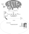
Mitochrondrial regulation of apoptosis. Bcl-2 family members regulate apoptosis by modulating the release of cytochrome c. Bax and Bak are pro-apoptotic. They physically interact and form channels that are permeable to cytochrome c. BH3-only members (e.g., Bid, Noxa, Puma) are pro-apoptotic and can modulate the conformation of Bax. Bcl-2 and Bcl-XL are anti-apoptotic and can block the function of Bax/Bak. The permeability transition pore (PTP), formed by the interaction of the adenine nucleotide translocator (ANT) and the voltage-dependent anion channel (VDAC) during the process of swelling, is a transmembrane channel that emerges at contact sites between the inner mitochondrial and the OMMs. The PTP has a role in regulating mitochondrial membrane potential and the release of cytochrome c. In the cytosol, cytochrome c, Apaf1, and procaspase-9 interact to form the apoptosome that drives the activation of caspase-3. The family of inhibitors of apoptosis (IAPs) blocks this process. The IAPs are inhibited by mitochondrially derived Smac, Diablo, and Omi. Caspase-3 cleaves many substrate proteins, some of which are endonucleases that translocate to the nucleus to cleave DNA into internucleosomal fragments (see DNA gel at lower right, showing molecular weight standards [M] in base pairs, developing rat cerebral cortex showing very low DNA fragmentation [lane 1], and developing rat brainstem inferior colliculus undergoing considerable apoptosis [lane 2]). Aif and endonuclease G are mitochondrially released proteins with nuclease activity that can translocate to the nucleus. Genomic DNA (double helix in nucleus) is the site of action of nucleases that induce strand nicks (X in helix). See text for detailed descriptions.
Fig 4
Fig 4
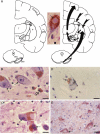
Brain atrophy and neurodegeneration in AD. Midsagittal views (center pictures) of the brains from an 85-year-old individual with AD and an 86-year-old normal individual. The microscopic neuropathological hallmarks of AD are senile plaques (scale bar=200μm), and neurofibrillary tangles (NFTs, scale bar=50μm) The silver stain detects deposits and accumulations of abnormal proteins such as a amyloid beta protein-containing senile plaques (open arrows) in cerebral cortex (top left image) and NFTs in neurons and neuronal tombstones (open arrows, top middle image). Antibodies can be used to detect protein constituents of NFTs in neurons such a hyperphosphorylated tau (open arrows in top right image). These abnormalities are microscopic pathological entities associated with AD.
Fig 5
Fig 5
Basal ganglia circuits in control of movement and SNpc neuron degeneration in people with PD. (A) The basal ganglia are comprised (left panel) of the caudate nucleus (CN), putamen (P), globus pallidus external (GPe) and internal (GPi) divisions, the subthalamic nucleus (STN), and the substantia nigra compact (SNpc) and reticular (SNr) divisions. The cerebral cortex and thalamus (T), although not part of the basal ganglia, participate in the connectivity loops (right panel). The major excitatory input to the striatum (the caudate nucleus and putamen) is from the cerebral cortex (top, right panel). The striatum, in turn, projects to the globus pallidus and the substantia nigra reticular division. Striatal activity is modulated by an extensive dopaminergic input from the SNpc. The major output of the basal ganglia is directed toward the thalamus, originating from GPi and from the substantia nigra reticular division (not shown). The thalamic projection to the cerebral cortex (premotor and supplementary motor areas) drives the activity of motor cortex that executes somatic movements. Between the two panels is a SNpc neuron with a LB (arrow) seen by hematoxylin–eosin (H&E) staining. (B and C) Degeneration of SNpc neurons in human PD. H&E staining shows that the degeneration of pigmented SNpc neurons is characterized by chromatolysis (B, hatched arrows) and nuclear condensation (C, hatched arrow), and severe soma attrition (C, inset hatched arrow). The neuronal chromatolysis (in B) is indicated by the eccentrically placed nucleus, pale cytoplasm, and peripheral margination of the Nissl substance. Glial/macrophage-like cells (B, open arrow) are laden with phagocytosed cellular debris. The nucleus of SNpc neurons undergoes considerable condensation (C, hatched arrow) while the Nissl substance dissipates, but before appreciable somal shrinkage. The cell body of SNpc neurons then becomes attritional (C, inset, hatched arrow), resulting in residual neurons that are ≈10–20% their normal size. The cell shown is an atrophic neuron, rather than a debris-laden macrophage, because of the presence of a condensed nucleus with a single prominent nucleolus. This degeneration pattern could be indicative of autophagy. (D) The nuclear condensation stage of pigmented SNpc neuron degeneration is characterized by the appearance of DNA double-strand breaks as detected by TUNEL (arrow, brown staining). (E) SNpc neurons accumulate cleaved caspase-3 (arrows, brown staining). Scale bars: B, 20μm (same for C and D); C inset, 6μm; E, 45μm.
Fig 6
Fig 6
Motor neurons in spinal cord degenerate in ALS. (A) In normal control individuals, the anterior horns of the spinal cord contain many large, multipolar motor neurons (dark cells). (B) In ALS cases, the anterior horn is depleted of large neurons (dark cells) and remaining neurons are atrophic. These attritional chromatolytic motor neurons display a dark condensed nucleus as seen microscopically. Scale bar in A=76μm (same for B). (C, D, and E) Nissl staining shows that the degeneration of motor neurons in familial ALS is characterized by shrinkage and progressive condensation of the cytoplasm and nucleus. The motor neuron in (C) (arrow) appears normal. It has a large, multipolar cell body and a large nucleus containing a reticular network of chromatin and a large nucleolus. Scale bar=7 μm (same for D and E).The motor neuron in (D) (arrow) has undergone severe somatodentritic attrition. The motor neuron in (E) is at near end-stage degeneration (arrow). The cell has shrunken to ≈10% of its normal size and has become highly condensed. The cell in (E) is identified as a residual motor neuron based on the nucleus and nucleolus (seen as eccentrically placed darkly stained component to the lower left of cytoplasmic pale area) and residual large Nissl bodies. (F) Cell death assays (e.g., TUNEL) identify subsets of motor neurons in the process of DNA fragmentation. Nuclear DNA fragmentation (brown labeling) occurs in motor neurons as the nucleus condenses and the cell body shrinks. Motor neurons in the somatodendritic attrition stage accumulate DNA double strand breaks. Scale bar=7 μm. (G) In individuals with ALS, p53 accumulates in the nucleus (brown labeling) of motor neurons. Scale bar=5 μm. (H) Degenerating motor neurons in human ALS are immunopositive for cleaved caspase-3 (black-dark green labeling) in the somatodendritic attrition stage. Around the nucleus (pale circle), motor neurons accumulate discrete mitochondria (brown-orange labeling, detected with antibody to cytochrome c oxidase subunit I) exhibiting little light microscopic evidence for swelling. Scale bar=5μm.

Similar articles

Cited by

References

    1. Zorov DB, Isave NK, Plotnikov EY, Zorova LD, Stelmashook EV, Vasileva AK, et al. The mitochondrion as Janus Bifrons. Biochemistry (Mosc) 2007;72:1115–26. - PubMed
    1. Halliwell B. Role of free radicals in the neurodegenerative diseases. Drugs Aging. 2001;18:685–716. - PubMed
    1. Nicholls DG. Mitochondrial function and dysfunction in the cell: its relevance to aging and aging-related disease. Int J Biochem Cell Biol. 2002;34:1372–81. - PubMed
    1. Wallace DC. A mitochondrial paradigm of metabolic and degenerative diseases, aging, and cancer: a dawn of evolutionary medicine. Annu Rev Genet. 2002;39:359–407. - PMC - PubMed
    1. Fridovich I. Superoxide radical and superoxide dismutases. Annu Rev Biochem. 1995;64:97–112. - PubMed

Publication types