. 2012 Apr 6;10(4):385-97.
doi: 10.1016/j.stem.2012.01.018.

Generation of multipotent lung and airway progenitors from mouse ESCs and patient-specific cystic fibrosis iPSCs

Affiliations

Generation of multipotent lung and airway progenitors from mouse ESCs and patient-specific cystic fibrosis iPSCs

Hongmei Mou et al. Cell Stem Cell. .

Erratum in

  • Cell Stem Cell. 2012 May 4;10(5):635

Abstract

Deriving lung progenitors from patient-specific pluripotent cells is a key step in producing differentiated lung epithelium for disease modeling and transplantation. By mimicking the signaling events that occur during mouse lung development, we generated murine lung progenitors in a series of discrete steps. Definitive endoderm derived from mouse embryonic stem cells (ESCs) was converted into foregut endoderm, then into replicating Nkx2.1+ lung endoderm, and finally into multipotent embryonic lung progenitor and airway progenitor cells. We demonstrated that precisely-timed BMP, FGF, and WNT signaling are required for NKX2.1 induction. Mouse ESC-derived Nkx2.1+ progenitor cells formed respiratory epithelium (tracheospheres) when transplanted subcutaneously into mice. We then adapted this strategy to produce disease-specific lung progenitor cells from human Cystic Fibrosis induced pluripotent stem cells (iPSCs), creating a platform for dissecting human lung disease. These disease-specific human lung progenitors formed respiratory epithelium when subcutaneously engrafted into immunodeficient mice.


Figures

Figure 1
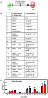
Figure 1. Anteriorization of Endoderm to Foregut Endoderm Promotes Nkx2.1+ Cell Differentiation
(A) Administration of 20 ng/ml BMP4, 20 ng/ml FGF2, and 5 nM GSK3iXV to definitive endoderm resulted in hindgut differentiation with CDX2 expression, and minimal NKX2.1 induction. Scale bar, 50 μm. (B) Definitive endoderm was treated with different combinations of BMP4/TGFβ agonists and antagonists as listed in the table for 2 days. The Foxa2+/Sox2+ cells were quantified as a percentage of positive cells out of the total number of Foxa2+ cells. The cells were further treated with 20 ng/ml BMP4, 20 ng/ml FGF2, and 5 nM GSK3iXV to induce NKX2.1 expression. The percentage of Nkx2.1+ cells was quantified as a percentage of the total cells present. All data were averaged from three independent experiments. (C) Representative immunofluorescence staining showing Foxa2+ and Sox2+ double positive cells after 2 days of treatment with 500 nM A-83-01. Scale bar, 100 μm. (D) Representative immunofluorescence staining showing NKX2.1 induction from A-83-01 treated endoderm cells after BMP4/FGF2/GSK3iXV treatment for 2 days. Scale bar, 100 μm.
Figure 2
Figure 2. ESC-Derived Nkx2.1+ Cells Are Devoid of Neuronal and Thyroid Markers, Are Proliferative, and Possess Markers of Proximal and Distal Lung Endoderm
(A) Immunofluorescence staining for FOXA2, TUJ1, and PAX8 showing that Nkx2.1+ cells are positive for endodermal marker FOXA2, negative for neuro-ectodermal marker TUJ1 (white arrow), and negative for thyroid marker PAX8. Scale bar, 50 μm. (B) Nkx2.1+ cells were proliferative as demonstrated by costaining for KI67. Scale bar, 100 μm. (C) Representative immunofluorescence staining showing subpopulations of Nkx2.1+ cells are positive for SOX2 (an airway progenitor marker) and FOXP2/SOX9 (multipotent lung progenitor marker). Scale bar, 100 μm.
Figure 3
Figure 3. BMP4, FGF2, and WNT Signaling Are Necessary for Lung Specification from Foregut Endoderm Cells
(A) Schematic strategy and time frame to generate Nkx2.1+ cells from foregut endoderm cells. (B) NKX2.1 induction using various combinations of BMP4, FGF2, WNT, and their antagonists. (C) NKX2.1 percentage was scored as the number of Nkx2.1+ cells out of the total cell number, average of three independent experiments.
Figure 4
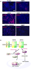
Figure 4. Smad-Dependent BMP Signaling Is Required for Lung Differentiation
(A) NKX2.1 induction with 20 ng/ml FGF2 and 5 nM GSK3iXV only (control) or with additional BMP4 (20 ng/ml), BMP2 (20 ng/ml), BMP7 (20 ng/ml), BMP4 (20 ng/ml) + 10 μM Dorsomorphin, or BMP4 (20 ng/ml) + 1 μM PD98059. Scale bar, 100 μm. (B) BMP signaling through SMAD-dependent pathways is required for lung Nkx2.1+ differentiation. In contrast, signaling through the MAP kinase pathway is not required for Nkx2.1 expression. Dorsomorphin inhibits BMP signaling through the SMAD pathway while PD98059 inhibits MAPK signaling.
Figure 5
Figure 5. Generation of Embryonic Airway Progenitors from Multipotent Lung Endoderm Cells
(A) Schematic strategy and time line to generate embryonic airway progenitors from multipotent lung endoderm cells. (B) Summary of signaling switches that are distinct in the proximal airway and the distal lung bud tip during the pseudoglandular stage of lung development. (C) Left panel: NKX2.1 and SOX2 staining after treatment of Nkx2.1+ lung endoderm cells at D9 with medium containing RA-supplemented B27, 20 ng/ml BMP7, 20 ng/ml FGF7, 100 nM IWR-1 (WNT antagonist), and 100 ng/ml Noggin for 2 days. Right panel: NKX2.1 and SOX2 staining after treatment of Nkx2.1+ lung endoderm cells at D9 with medium containing RA-supplemented B27, 20 ng/ml BMP7, 20 ng/ml FGF7, 100 nM IWR-1 (WNT antagonist), and 1 μM PD98059 for 2 days. Scale bar, 50 μm. (D) Immunofluorescence staining showing a subpopulation of Nkx2.1+ cells positive for P63 (closed arrows). Scale bar, 25 μm.
Figure 6
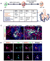
Figure 6. Differentiation of Airway Epithelium from Mouse-ESC-Derived Lung Endoderm upon Subcutaneous Engraftment
(A) Schematic strategy and time line to differentiate ESC-derived embryonic airway progenitors. (B) Immunofluorescence staining showing that some Nkx2.1+ cells are positive for SOX2. Scale bar, 100 μm. (C) Immunofluorescence staining showing differentiation of ESC-derived Nkx2.1+ cells into p63+ airway basal stem cells, CC10+ Clara cells, FoxJ1+ ciliated cells, and Muc5ac+ goblet cells. The arrow points to a representative positive cell for each category. Scale bar, 100 μm. (D) Confocal imaging showing differentiation of various airway epithelial cells. Scale bar, 100 μm.
Figure 7
Figure 7. Stepwise Differentiation of Nkx2.1+ Lung Progenitors from Human iPSCs
(A) Schematic strategy and time line to generate Nkx2.1+ lung multipotent progenitors from human iPSCs. (B) High yield of definitive endoderm from CF1 RiPSCs was obtained after treatment for 4 days in RPMI-1640 medium in the presence of 2% B27 supplement, Activin A (100 ng/ml), and 5 μM PI3 Kinase inhibitor LY294002 with more than 90% of cells coexpressing transcription factors SOX17 and FOXA2. Scale bar, 200 μm. (C) Anteriorization of endoderm into foregut endoderm cells with SOX2 expression in Foxa2+ cells derived from CF1 RiPSCs after 4 days of treatment with 500 nM A-83-01 (TGFβ antagonist) and 100 ng/ml Noggin (BMP4 antagonist). Scale bar, 100 μm. (D) Left panel: NKX2.1 staining after anteriorization to foregut cells at D8 with serum-free medium containing 20 ng/ml BMP4, 20 ng/ml FGF2, and 5 nM GSK3iXV for 4 days. Scale bar, 100 μm. Right panel: costaining of Nkx2.1-positive cells with SOX2, SOX9, TUJ1 (arrows), and PAX8 demonstrated a lack of thyroid and neuronal differentiation and the presence of multipotent distal tip progenitors and Nkx2.1+/Sox2+ airway progenitors. Scale bar, 50 μm. (E). Confocal image after immunofluorescence staining showing that some Nkx2.1+ spheres contain basal cells positive for p63. Scale bar, 40 μm.

Similar articles

Cited by

References

    1. Accurso FJ, Rowe SM, Clancy JP, Boyle MP, Dunitz JM, Durie PR, Sagel SD, Hornick DB, Konstan MW, Donaldson SH, et al. Effect of VX-770 in persons with cystic fibrosis and the G551D-CFTR mutation. N Engl J Med. 2010;363:1991–2003. - PMC - PubMed
    1. Ameri J, Ståhlberg A, Pedersen J, Johansson JK, Johannesson MM, Artner I, Semb H. FGF2 specifies hESC-derived definitive endoderm into foregut/midgut cell lineages in a concentration-dependent manner. Stem Cells. 2010;28:45–56. - PubMed
    1. Aubin J, Davy A, Soriano P. In vivo convergence of BMP and MAPK signaling pathways: impact of differential Smad1 phosphorylation on development and homeostasis. Genes Dev. 2004;18:1482–1494. - PMC - PubMed
    1. Bellusci S, Henderson R, Winnier G, Oikawa T, Hogan BL. Evidence from normal expression and targeted misexpression that bone morphogenetic protein (Bmp-4) plays a role in mouse embryonic lung morphogenesis. Development. 1996;122:1693–1702. - PubMed
    1. Cao L, Gibson JD, Miyamoto S, Sail V, Verma R, Rosenberg DW, Nelson CE, Giardina C. Intestinal lineage commitment of embryonic stem cells. Differentiation. 2011;81:1–10. - PubMed

Publication types

MeSH terms