. 2012 Apr 6;10(4):398-411.
doi: 10.1016/j.stem.2012.01.019.

Efficient derivation of purified lung and thyroid progenitors from embryonic stem cells

Affiliations

Efficient derivation of purified lung and thyroid progenitors from embryonic stem cells

Tyler A Longmire et al. Cell Stem Cell. .

Abstract

Two populations of Nkx2-1(+) progenitors in the developing foregut endoderm give rise to the entire postnatal lung and thyroid epithelium, but little is known about these cells because they are difficult to isolate in a pure form. We demonstrate here the purification and directed differentiation of primordial lung and thyroid progenitors derived from mouse embryonic stem cells (ESCs). Inhibition of TGFβ and BMP signaling, followed by combinatorial stimulation of BMP and FGF signaling, can specify these cells efficiently from definitive endodermal precursors. When derived using Nkx2-1(GFP) knockin reporter ESCs, these progenitors can be purified for expansion in culture and have a transcriptome that overlaps with developing lung epithelium. Upon induction, they can express a broad repertoire of markers indicative of lung and thyroid lineages and can recellularize a 3D lung tissue scaffold. Thus, we have derived a pure population of progenitors able to recapitulate the developmental milestones of lung/thyroid development.


Figures

Figure 1
Figure 1. Inhibition of BMP and TGFβ signaling alters endodermal patterning and modulates competence of ESCs to differentiate into either endodermal Nkx2-1+ or neuronal Nkx2-1+ cells
(A) Schematic of AP patterning of the endodermal gut tube in the developing embryo (~E8.5-9). Domains of expression of genes that demarcate prospective organ fields. (B) Kinetics of expression of knock-in reporters, Foxa2-hCD4 and Foxa3-hCD25, indicating effect of Activin stimulation alone (left panel) compared to Activin followed by exposure to the BMP and TGFβ inhibitors, Noggin and SB431542 (Nog/SB for 24 hours. (C) Nkx2-1GFP knock-in mouse embryo and lung whole mount (both at E14.5) with GFP fluorescence visible in the forebrain, thyroid and lung (thyroid in figure S2). In the lung and trachea, fluorescence appears in an epithelial pattern (Tr; arrowhead). (D) Effect of Nog/SB exposure on the competence of day 5 cells to specify to Nkx2-1+ endoderm by day 14. Representative flow cytometry dot plots and summary bar graph indicate percentages of GFP+ cells in each condition (n=4; avg±SEM; *p<0.001). qRT-PCR analysis of indicated genes comparing day 14 sorted GFP+ to GFP- cells (bars=avg fold change in gene expression±SD). (E) Effect of WFKBE with vs without FGF2 supplementation on GFP+ cell morphology. GFP fluorescence and phase contrast overlay images are shown for D12 (pre-sort) ESC-derived GFP+ cells, which occur only rarely in the absence of FGF2 (top panel) and express neuroectodermal markers, such as Pax6 (data not shown). In contrast, GFP+ outgrowth cells in presence of FGF2 (lower panel) show a different morphology and are endodermal (see markers in panel D). Size bar=100μm. (F) Prolonged Nog/SB exposure specifies Nkx2-1+ neuronal cells by day 13, as indicated by flow cytometry and kinetics of induction of neuronal markers (Olig2 and Tuj1), loss of endodermal marker, Foxa2, and loss of early thyroid marker, Pax8. [see also figures S1-S4].
Figure 2
Figure 2. Purified endodermal Nkx2-1GFP+ progenitors derived from ESCs proliferate in culture and express a repertoire of lung and thyroid lineage genes
(A) Schematic of culture protocol for directed differentiation of ESCs into Nkx2-1GFP+ (B) Sort gate used to purify GFP+ cells for replating and outgrowth. (C) Expression of Nkx2-1 mRNA and indicated marker genes (D) for each cell population, quantified by real time RT-PCR. E18.5 lung and thyroid RNA extracts served as positive controls. Bars indicate average fold change in gene expression over ESCs±SEM (n=3 independent experiments). DCI+K=cells exposed to lung maturation media from D22-25 [see also figure S5].
Figure 3
Figure 3. Purified ESC-derived populations expressing Nkx2-1 protein co-express markers of early developing endoderm
(A) Heterogeneous ESC-derived cultures on day 14 (D14; 1 day before GFP+ cell sorting) immunostained for Nkx2-1, Foxa2, Sox2, Sox9, and Tuj1 proteins, demonstrate co-expression of Foxa2 in the majority of Nkx2-1+ cells and expression of Sox2 or Sox9 in subsets of Nx2-1+ cells and in Nkx2-1- cells. In contrast, Tuj1+ cells do not overlap with Nkx2-1+ cells. In these wells ~16% of all cells were Nkx2-1GFP+ by flow cytometry. (B) After purification of the Nkx2-1GFP+ cells shown in A by flow cytometry on D15, outgrowth cells stained on D24 for each indicated marker show more clearly defined morphology than before sorting in panel A. Arrowhead= rare neuronal (Tuj1+) cell found after cell sorting. At this D24 time point ~60% of cells retained Nkx2-1GFP expression by flow cytometry. Size bar=100 μm. Nuclei counterstained with DAPI (blue).
Figure 4
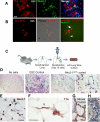
Figure 4. Alveolar differentiation repertoire of ESC-derived Nkx2.1+ lung progenitors
(A,B) Immunostaining for alveolar epithelial markers T1α, pro-SPC and Nkx2-1 on cells at the completion of the 25 day directed differentiation protocol. ESCs sorted on day 15 based on Nkx2-1GFP+ expression subsequently gave rise to (A) cells reminiscent of type 1 alveolar epithelial cells (AEC1) as they lost Nkx2.1 nuclear protein expression (green immunostain) while expressing T1α protein. (B) Other patches of cells appeared more reminiscent of distal SPC+ alveolar epithelial cells as they expressed punctate cytoplasmic pro-SPC protein and displayed SPC promoter activation while retaining Nkx2-1GFP expression. Arrow=SPC-dsRed and Nkx2-1GFP co-expressing cell (orange). Arrowhead= cell expressing only Nkx2-1GFP. (C) Schematic summarizing the decellularization-recellularization assay. (D) H&E stains of lung sections showing lung scaffold appearance with no recellularization (left panel) vs hypercellular sheets following reseeding with undifferentiated ESCs (middle panel) vs cells of alveolar structural morphologies after seeding with Nkx2-1GFP+ purified ESC-derived progenitors(right panel). Size bars=100μm in three left panels. Zoom of the indicated boxed region is shown in far right panel with size bar=20μm. (E) Nkx2-1+ nuclear protein (brown; arrowheads) immunostaining of engrafted cuboidal epithelial cells in the corners of alveoli, derived 10 days after re-seeding with Nkx2.1GFP+ sorted cells. Arrow= flattened nucleus of an Nkx2.1 negative cell (purple) lining the alveolar septum. Many of these flattened cells were T1α+ (F; arrowhead) Size bars=20μm. (G) Control mouse lung without decellularization showing T1α apical membrane staining pattern (brown) of mature AEC1 [see also figure S7]. (H) Ciliated airway epithelial cell (arrow) 10 days after re-seeding with differentiated/unsorted ESC-derived cells. Size bar=20μm. All nuclei counterstained with hematoxylin (purple). [See also supplemental figures S6 and S7].
Figure 5
Figure 5. Combinatorial FGF and BMP signaling promotes efficient derivation of proliferating Nkx2-1GFP+ ESC-derived cells
(A) Percentage of Nkx2-1GFP+ cells derived from ESCs on D14 when individual growth factors were withdrawn from the 6 factor (6F) ventralizingWFKBE+FGF2 cocktail. two-tailed t test *p<0.05. (B) BrdU labeling index (after 24 hours vs 0 hours of BrdU exposure) measured by flow cytometry in each condition from panel A. Cells are scored as BrdU+ based on histogram gates set on cells that did not receive BrdU. (C) Immunostaining for Nkx2-1 and Ki67 nuclear proteins in D15 ESC-derived cells. (D) Model of directed differentiation of ESCs into neuroectodermal Nkx2-1+ cells vs endodermal Nkx2-1+ cells with lung and thyroid differentiation potential.
Figure 6
Figure 6. Epigenetic changes in the Nkx2-1 and Oct4 proximal promoter regions during directed differentiation of ESCs
(A) Schematic of cell fate decision in ESC-derived endodermal cells to commit to an Nkx2-1 descendant lineage. (B) Kinetics of Nkx2-1GFP expression during 22 days of ESC directed differentiation, following the protocol shown in figure 2. (C) Chromatin immunoprecipitation and bisulfite sequencing studies demonstrate the DNA methylation and histone trimethylation states of the proximal promoters of the Nkx2-1 and Oct4 loci in each cell population during development. Arrows and numbers indicate PCR primer binding sites relative to the ATG start site. Open circles=unmethylated DNA CpG sites, closed circles=methylated CpG sites. Lung Nkx2-1GFP+ vs Nkx2-1GFP- samples were prepared by sorting primary lung cell digests from a 3 week old mouse. MLE-15= mouse lung epithelial cell line.
Figure 7
Figure 7. Differentially expressed genes in ESC-derived Nkx2-1GFP+ vs Nkx2-1GFP- cells
(A) Heat map of 1267 genes differentially expressed between triplicate samples of Nkx2-1GFP+ vs Nkx2-1GFP- ESC-derived cells, sorted on day 14 of differentiation. (B) Gene Ontology analysis of the 594 genes upregulated in GFP+ cells (DAVID on-line analysis). (C) Selections of the 1267 differentially expressed genes demonstrate differing gene programs relating to thyroid lineage, epithelial signaling and mesenchymal programs, or (D) genes whose promoters are known targets of Nkx2-1 protein binding in E11.5 embryonic lung epithelial cells in vivo. Gene.symb=NCBI gene ID. LogFC=log2 fold change in gene expression. [see also table S1]

References

    1. Ali NN, Edgar AJ, Samadikuchaksaraei A, Timson CM, Romanska HM, Polak JM, Bishop AE. Derivation of type II alveolar epithelial cells from murine embryonic stem cells. Tissue Eng. 2002;8:541–550. - PubMed
    1. Ameri J, Ståhlberg A, Pedersen J, Johansson JK, Johannesson MM, Artner I, Semb H. FGF2 specifies hESC-derived definitive endoderm into foregut/midgut cell lineages in a concentration-dependent manner. Stem Cells. 2010;28:45–56. - PubMed
    1. Arufe MC, Lu M, Kubo A, Keller G, Davies TF, Lin RY. Directed differentiation of mouse embryonic stem cells into thyroid follicular cells. Endocrinology. 2006;147:3007–3015. - PMC - PubMed
    1. Arufe MC, Lu M, Lin RY. Differentiation of murine embryonic stem cells to thyrocytes requires insulin and insulin-like growth factor-1. Biochem Biophys Res Commun. 2009;381:264–270. - PMC - PubMed
    1. Cao Y, Vo T, Millien G, Tagne JB, Kotton D, Mason RJ, Williams MC, Ramirez MI. Epigenetic mechanisms modulate thyroid transcription factor 1-mediated transcription of the surfactant protein B gene. J Biol Chem. 2010;285:2152–2164. - PMC - PubMed

Publication types

Substances

Associated data