Review
. 2012 Apr;2(2):128-33.
doi: 10.1016/j.coviro.2012.01.005. Epub 2012 Feb 16.

M2 protein from influenza A: from multiple structures to biophysical and functional insights

Affiliations
Review

M2 protein from influenza A: from multiple structures to biophysical and functional insights

Timothy A Cross et al. Curr Opin Virol. 2012 Apr.

Abstract

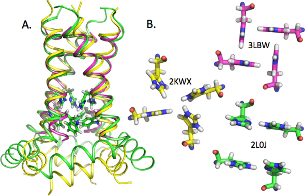
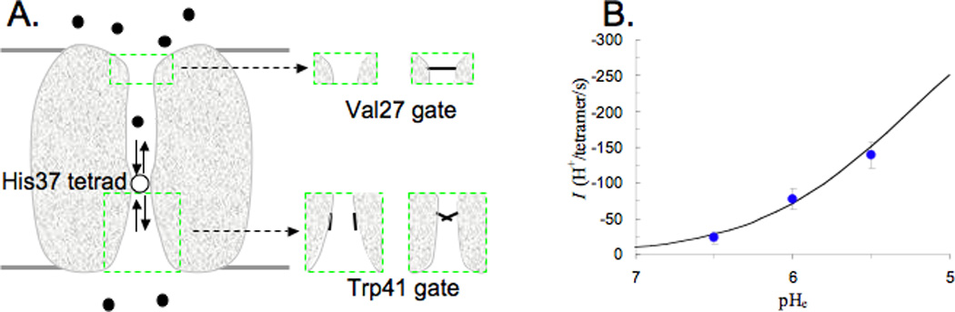
The M2 protein from influenza A is a proton channel as a tetramer, with a single transmembrane helix from each monomer lining the pore. Val27 and Trp41 form gates at either end of the pore and His37 mediates the shuttling of protons across a central barrier between the N-terminal and C-terminal aqueous pore regions. Numerous structures of this transmembrane domain and of a longer construct that includes an amphipathic helix are now in the Protein Data Bank. Many structural differences are apparent from samples obtained in a variety of membrane mimetic environments. High-resolution structural results in lipid bilayers have provided novel insights into the functional mechanism of the unique HxxxW cluster in the M2 proton channel.


Figures

Fig. 1
Fig. 1
Comparison of three recent structures of M2, shown with the backbone as a helical ribbon and the His37 and Trp41 sidechains as sticks. A) 2L0J was obtained by solid state NMR spectroscopy on the M2 conductance domain (residues 22–62) in liquid crystalline lipid bilayers at pH 7.5 (green). 2KWX was obtained by solution NMR spectroscopy on the conductance domain (residues 18–60) in detergent micelles at pH 7.5 (yellow). 3LBW was obtained by x-ray crystallography on the TM domain (residues 22–46) in an octylglucoside environment at pH 6.5 (magneta). The ssNMR and x-ray structures of the TM helices superimpose very well, with a slight deviation at the C-terminus. The tilt of the solution NMR TM helix is significantly less than in the other two structures. Relative to the ssNMR structure, the amphipathic helices in the solution NMR structure have a different location with regard to both depth in the ‘membrane’ environment and lateral position. The His37 and Trp41 sidechains are shown for 2L0J. B) Comparison of the histidine sidechains in the three M2 structures displayed in A) as viewed from the viral interior (amphipathic helix side). The His37 sidechain torsion angles for 2KWX are similar to those for 2L0J, but the imidazole-imidzolium hydrogen bonds are not formed. As such the His37 sidechain conformations in 2KWX appear to be unstable due to charge repulsion. The His37 sidechain conformations in 3LBW provide an alternative, but more limited mechanism through cation-π interactions for charge dispersion and for structural stabilization.[22]
Fig. 2
Fig. 2
Mathematical model for calculating the rate of proton transport. A) Illustration of the model. A permeant proton, starting from the exterior bulk solution, diffuses toward the N-terminal pore, passes through the Val27 gate, and binds to the +2 charged His37 tetrad. A proton then dissociates from the His37 tetrad, either back to the N-terminal pore, or into the C-terminal pore, from which it passes by the Trp41 gate and diffuses into the interior bulk solution. The insets illustrate the fluctuating Val27 and Trp41 gates. B) Comparison of calculated (curve) and measured (symbols) rates of proton transport as functions of the exterior pH, pHe. The experimental data are from Ref. (the membrane potential was -114 mV and the interior pH was 8), In the calculations, the rate constant for binding to the His37 tetrad for a proton from the interior side is 1.2 × 10⁸ M−1s−1, the rate constant for releasing the proton from the His37 tetrad to the interior side is 3.6 × 10³s−1, and the probability for the Trp41 gate to be open is ~5%.

References

    1. Pinto LH, Lamb RA. Influenza virus proton channels. Photochem Photobiol Sci. 2006;5:629–632. - PubMed
    1. Pinto LH, Holsinger LJ, Lamb RA. Influenza virus M2 protein has ion channel activity. Cell. 1992;69:517–528. - PubMed
    1. Wang J, Qiu JX, Soto C, DeGrado WF. Structural and dynamic mechanisms for the function and inhibition of the M2 proton channel from influenza A virus. Curr Opin Struct Biol. 2011;21:68–80. - PMC - PubMed
    1. Balannik V, Wang J, Ohigashi Y, Jing X, Magavern E, Lamb RA, Degrado WF, Pinto LH. Design and pharmacological characterization of inhibitors of amantadine-resistant mutants of the M2 ion channel of influenza A virus. Biochemistry. 2009;48:11872–11882. - PMC - PubMed
    1. Anfinsen CB. Principles that govern the folding of protein chains. Science. 1973;181:223–230. - PubMed

Publication types

Substances