Review
. 2012 Apr;2(2):142-50.
doi: 10.1016/j.coviro.2012.02.003. Epub 2012 Mar 5.

TRIM5 structure, HIV-1 capsid recognition, and innate immune signaling

Affiliations
Review

TRIM5 structure, HIV-1 capsid recognition, and innate immune signaling

Markus G Grütter et al. Curr Opin Virol. 2012 Apr.

Abstract

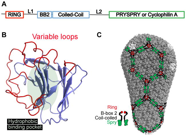
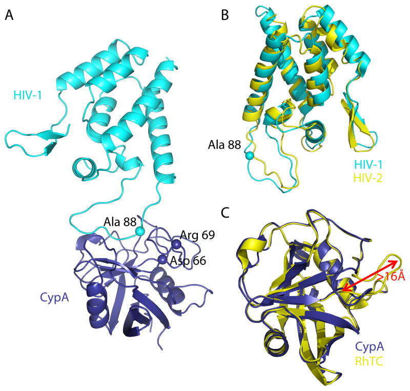
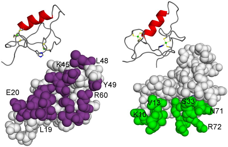
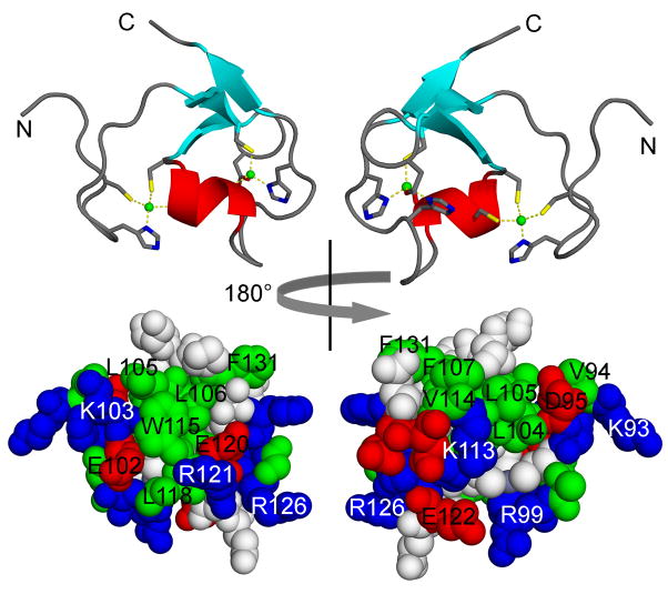
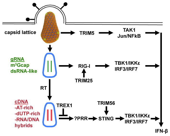
TRIM5 is a restriction factor that blocks retrovirus infection soon after the virion core enters the cell cytoplasm. Restriction activity is targeted to the virion core via recognition of the capsid protein lattice that encases the viral genomic RNA. In common with all of the many TRIM family members, TRIM5 has RING, B-box, and coiled-coil domains. As an E3 ubiquitin ligase TRIM5 cooperates with the heterodimeric E2, UBC13/UEV1A, to activate the TAK1 (MAP3K7) kinase, NF-κB and AP-1 signaling, and the transcription of inflammatory cytokines and chemokines. TAK1, UBC13, and UEV1A all contribute to TRIM5-mediated retrovirus restriction activity. Interaction of the carboxy-terminal PRYSPRY or cyclophilin domains of TRIM5 with the retroviral capsid lattice stimulates the formation of a complementary lattice by TRIM5, with greatly increased TRIM5 E3 activity, and host cell signal transduction. Structural and biochemical studies on TRIM5 have opened a much needed window on how the innate immune system detects the distinct molecular features of HIV-1 and other retroviruses.


Figures

Figure 1
Figure 1
Figure 1A: Schematic representation of the domains within TRIM5, with relative positions of the domains along the linear sequence indicated. RING, really interesting new gene; L1, linker 1; BB2, B box 2; L2, linker 2. Figure 1B. Ribbon diagram of a model of the human TRIM5α PRYSPRY (B30.2) domain. Variable loops extending from the β-sandwich are marked in red, the putative hydrophobic binding pocket is indicated by the light green oval. Fig. 1C. Model of the HIV-1 capsid fullerene cone with hexagonal TRIM5α lattice superimposed, illustrating a putative recognition mode of capsid by TRIM5α, as in reference [40]. The color code for the TRIM5α domains is indicated in the schematic model of the TRIM5α dimer at the bottom left of the panel.
Figure 2
Figure 2
Figure 2A: Cartoon representation of HIV-1-CypA complex (PDB 1AK4, Chains A and D) in which HIV-1 is in cyan and CypA is in dark blue. The proximity of HIV-1 CA residue Ala 88 to the CypA residues Asp 66 and Arg 69 (spheres) is shown. Figure 2B: Cartoon representation of the N-terminal capsid domains of HIV-1 (PWB 1GWP, first NMR model) in cyan and of HIV-2 (PDB 2WLV, chain A) in yellow after SSM superposition. The cyan sphere indicates the position of Ca Ala 88 in HIV-1, which is deleted in HIV-2. Figure 2C: Cartoon representation of CypA (PDB 2CPL) in dark blue SSM superimposed on RhTC (PDB 2WLW) in yellow. Loop 64–74, which contains amino acid replacements D66N and R69H has undergone a rearrangement of >16Å.
Figure 3
Figure 3
NMR solution structure of human TRIM5α, amino acid residues 1 to 78, encompassing the RING domain [56]. Top: ribbon diagrams with Zn2+ ions indicated as green balls. Coordinating residues are highlighted. Bottom: surface representations. Left: the RING-RING interaction region is indicated in magenta. Right: the E2 binding region is indicated in green.
Figure 4
Figure 4
NMR solution structure of human TRIM5α amino acid residues 86 to 131, encompassing the B-box 2 domain [58]. Top: ribbon diagrams with Zn2+ ions indicated as green balls, in a CHCDC2H2 coordination mode. Coordinating residues are highlighted. Bottom: surface representations. Left: hydrophobic surface cluster 1. Right: hydrophobic surface cluster 2.
Figure 5
Figure 5
Schematic diagram showing involvement in the innate immune response to HIV-1 of some of the many TRIM proteins. The role of TRIM5 has been confirmed, and, based on the evidence, the roles of TRIM25 and TRIM56 are likely. TRIM5 recognition of the capsid lattice stimulates its E3 ubiquitin ligase activity [39]. This activates TAK1 and downstream NF-κB and AP-1 factors. Such signalling is required for, but not sufficient to activate, interferon-beta transcription. TRIM25 is required for signaling by the RIG-I pattern recognition receptor [81,82]. RIG-I responds to the highly, structured HIV-1 genomic RNA [79,80]. TRIM56 is required for STING-dependent signalling from the cytoplasmic pattern recognition receptor for DNA [75], that is activated when HIV-1 infects TREX1-deficient cells [74].

References

    1. Reymond A, Meroni G, Fantozzi A, Merla G, Cairo S, Luzi L, Riganelli D, Zanaria E, Messali S, Cainarca S, et al. The tripartite motif family identifies cell compartments. EMBO J. 2001;20:2140–2151. - PMC - PubMed
    1. Nisole S, Stoye JP, Saïb A. TRIM family proteins: retroviral restriction and antiviral defence. Nat Rev Microbiol. 2005;3:799–808. - PubMed
    1. Han K, Lou DI, Sawyer SL. Identification of a Genomic Reservoir for New TRIM Genes in Primate Genomes. PLoS Genet. 2011;7:e1002388. Identification of new human TRIM genes, bringing total number to nearly 100. - PMC - PubMed
    1. Uchil PD, Quinlan BD, Chan W-T, Luna JM, Mothes W. TRIM E3 ligases interfere with early and late stages of the retroviral life cycle. PLoS Pathog. 2008;4:e16. Demonstration that many TRIM genes restrict retroviruses. - PMC - PubMed
    1. Matloubian M, Concepcion RJ, Ahmed R. CD4+ T cells are required to sustain CD8+ cytotoxic T-cell responses during chronic viral infection. J Virol. 1994;68:8056–8063. - PMC - PubMed

Publication types