. 2012:110:325-66.
doi: 10.1016/B978-0-12-388403-9.00013-8.

Multi-scale modeling of tissues using CompuCell3D

Affiliations

Multi-scale modeling of tissues using CompuCell3D

Maciej H Swat et al. Methods Cell Biol. 2012.

Abstract

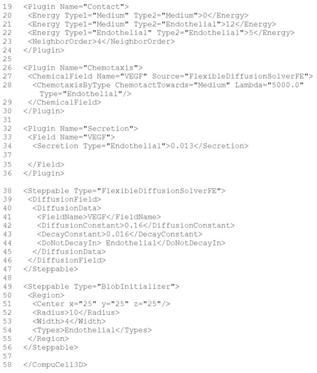
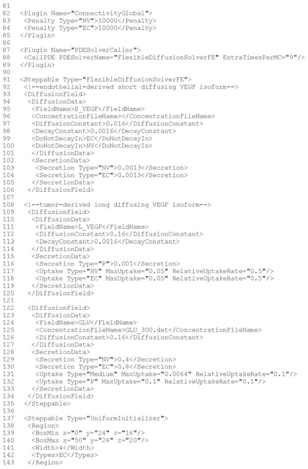
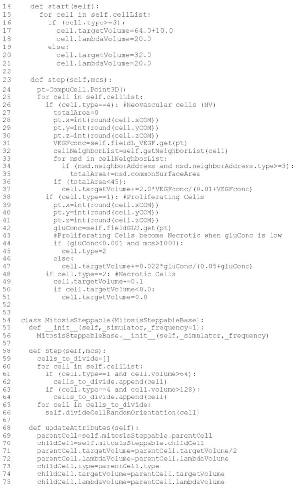
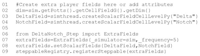
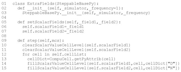
The study of how cells interact to produce tissue development, homeostasis, or diseases was, until recently, almost purely experimental. Now, multi-cell computer simulation methods, ranging from relatively simple cellular automata to complex immersed-boundary and finite-element mechanistic models, allow in silico study of multi-cell phenomena at the tissue scale based on biologically observed cell behaviors and interactions such as movement, adhesion, growth, death, mitosis, secretion of chemicals, chemotaxis, etc. This tutorial introduces the lattice-based Glazier-Graner-Hogeweg (GGH) Monte Carlo multi-cell modeling and the open-source GGH-based CompuCell3D simulation environment that allows rapid and intuitive modeling and simulation of cellular and multi-cellular behaviors in the context of tissue formation and subsequent dynamics. We also present a walkthrough of four biological models and their associated simulations that demonstrate the capabilities of the GGH and CompuCell3D.


Figures

Fig. 1
Fig. 1
GGH representation of an index-copy attempt for two cells on a 2D square cell lattice – The “white” pixel (source) attempts to replace the “grey” pixel (target). The probability of accepting the index copy is given by Eq. (2).
Fig. 2
Fig. 2
Flow chart of the GGH algorithm as implemented in CompuCell3D.
Fig. 3
Fig. 3
CellDraw graphics tools and GUI. (For color version of this figure, the reader is referred to the web version of this book.)
Fig. 4
Fig. 4
Invoking the CompuCell3D Simulation Wizard from Twedit++. (For color version of this figure, the reader is referred to the web version of this book.)
Fig. 5
Fig. 5
Specification of basic cell-sorting properties in Simulation Wizard. (For color version of this figure, the reader is referred to the web version of this book.)
Fig. 6
Fig. 6
Specification of cell-sorting cell types in Simulation Wizard. (For color version of this figure, the reader is referred to the web version of this book.)
Fig. 7
Fig. 7
Selection of cell-sorting cell behaviors in Simulation Wizard. (For color version of this figure, the reader is referred to the web version of this book.)
Fig. 8
Fig. 8
Snapshots of the cell-lattice configurations for the cell-sorting simulation in Listing 1. The boundary-energy hierarchy drives NonCondensing (light grey) cells to surround Condensing (dark grey) cells. The white background denotes surrounding Medium.
Fig. 9
Fig. 9
Specification of the angiogenesis chemical field in Simulation Wizard. (For color version of this figure, the reader is referred to the web version of this book.)
Fig. 10
Fig. 10
Specification of angiogenesis cell behaviors in Simulation Wizard. (For color version of this figure, the reader is referred to the web version of this book.)
Fig. 11
Fig. 11
Specification of angiogenesis secretion parameters in Simulation Wizard. (For color version of this figure, the reader is referred to the web version of this book.)
Fig. 12
Fig. 12
Specification of angiogenesis chemotaxis properties in Simulation Wizard. (For color version of this figure, the reader is referred to the web version of this book.)
Fig. 13
Fig. 13
An initial cluster of adhering endothelial cells forms a capillary-like network via sprouting angiogenesis. (A) 0 h (0 MCS); (B) ~2 h (100 MCS); (C) ~5 h (250 MCS); (D): ~18 h (1100 MCS). (For color version of this figure, the reader is referred to the web version of this book.)
Fig. 14
Fig. 14
Specification of vascular tumor chemical fields in the Simulation Wizard. (For color version of this figure, the reader is referred to the web version of this book.)
Fig. 15
Fig. 15
Specification of vascular tumor cell behaviors in Simulation Wizard. (For color version of this figure, the reader is referred to the web version of this book.)
Fig. 16
Fig. 16
Specification of vascular tumor chemotaxis properties in Simulation Wizard. (For color version of this figure, the reader is referred to the web version of this book.)
Fig. 17
Fig. 17
Two-dimensional snapshots of the vascular tumor simulation taken at: (A) 0 MCS; (B) 500 MCS; (C) 2000 MCS; (D) 5000 MCS. Red and yellow cells represent endothelial cells and neovascular endothelial cells, respectively. (See color plate.)
Fig. 18
Fig. 18
Diagram of Delta–Notch feedback regulation between and within cells.
Fig. 19
Fig. 19
Initial Notch (left) and Delta (right) concentrations in the Delta–Notch model. (For color version of this figure, the reader is referred to the web version of this book.)
Fig. 20
Fig. 20
Dynamics of the Notch concentrations of cells in the Delta–Notch model. Snapshots taken at 10, 100, 300, 400, 450, and 600 MCS. (See color plate.)
Listing 1
Listing 1
Simulation-Wizard-generated draft CC3DML (XML) code for cell sorting.
Listing 2
Listing 2
CC3DML code for the angiogenesis model.
Listing 2
Listing 2
CC3DML code for the angiogenesis model.
Listing 3
Listing 3
Simple Python loop.
Listing 4
Listing 4
Iterating over the inventory of CC3D cells in Python.
Listing 5
Listing 5
Sample CC3D steppable class.
Listing 6
Listing 6
The Main Python script initializes the vascular tumor simulation and runs the main simulation loop.
Listing 7
Listing 7
CC3DML specification of the vascular tumor model’s initial cell layout, PDE solvers, and key cellular behaviors.
Listing 7
Listing 7
CC3DML specification of the vascular tumor model’s initial cell layout, PDE solvers, and key cellular behaviors.
Listing 7
Listing 7
CC3DML specification of the vascular tumor model’s initial cell layout, PDE solvers, and key cellular behaviors.
Listing 7
Listing 7
CC3DML specification of the vascular tumor model’s initial cell layout, PDE solvers, and key cellular behaviors.
Listing 8
Listing 8
Vascular tumor model Python steppables. The VolumeParametersSteppable adjusts the properties of the cells in response to simulation events and the MitosisSteppable implements cell division.
Listing 8
Listing 8
Vascular tumor model Python steppables. The VolumeParametersSteppable adjusts the properties of the cells in response to simulation events and the MitosisSteppable implements cell division.
Listing 9
Listing 9
Jarnac specification of the Delta–Notch coupling model in Fig. 17.
Listing 10
Listing 10
Registering DeltaNotchClass in the main Python script, DeltaNotch.py in the Delta–Notch model.
Listing 11
Listing 11
Implementation of the _init_ and start functions of the DeltaNotchClass in the Delta–Notch model.
Listing 12
Listing 12
Implementation of a step function (continuation of the code from Listing 11) to calculate in the DeltaNotchClass in the Delta–Notch model.
Listing 13
Listing 13
Adding extra visualization fields in the main Python script DeltaNotch.py in the Delta–Notch model.
Listing 14
Listing 14
Steppable to visualize the concentrations of Delta and Notch in each cell in the Delta–Notch model.

References

    1. Alber MS, Jiang Y, Kiskowski MA. Lattice gas cellular automation model for rippling and aggregation in myxobacteria. Physica D. 2004;191:343–358.
    1. Alber MS, Kiskowski MA, Glazier JA, Jiang Y. On cellular automation approaches to modeling biological cells. In: Rosenthal J, Gilliam DS, editors. Mathematical Systems Theory in Biology, Communication and Finance. Springer-Verlag; New York: 2002. pp. 1–40.
    1. Alber M, Chen N, Glimm T, Lushnikov P. Multiscale dynamics of biological cells with chemotactic interactions: from a discrete stochastic model to a continuous description. Phys Rev E. 2006;73:051901. - PubMed
    1. Armstrong PB, Armstrong MT. A role for fibronectin in cell sorting out. J Cell Sci. 1984;69:179–197. - PubMed
    1. Armstrong PB, Parenti D. Cell sorting in the presence of cytochalasin B. J Cell Biol. 1972;55:542–553. - PMC - PubMed

Publication types

Substances

LinkOut - more resources