Tumefactive multiple sclerosis requiring emergent biopsy and histological investigation to confirm the diagnosis: a case report
- PMID: 22483341
- PMCID: PMC3337287
- DOI: 10.1186/1752-1947-6-104
Tumefactive multiple sclerosis requiring emergent biopsy and histological investigation to confirm the diagnosis: a case report
Abstract
Introduction: Tumefactive multiple sclerosis is a demyelinating disease that demonstrates tumor-like features on magnetic resonance imaging. Although diagnostic challenges without biopsy have been tried by employing radiological studies and cerebrospinal fluid examinations, histological investigation is still necessary for certain diagnosis in some complicated cases.
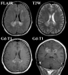
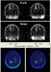
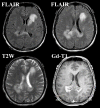
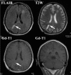
Case presentation: A 37-year-old Asian man complaining of mild left leg motor weakness visited our clinic. Magnetic resonance imaging demonstrated high-signal lesions in bilateral occipital forceps majors, the left caudate head, and the left semicentral ovale on fluid-attenuated inversion recovery and T2-weighted imaging, and these lesions were enhanced by gadolinium-dimeglumin. Tumefactive multiple sclerosis was suspected because the enhancement indistinctly extended along the corpus callosum on magnetic resonance imaging and scintigraphy showed a low malignancy of the lesions. But oligoclonal bands were not detected in cerebrospinal fluid. In a few days, his symptoms fulminantly deteriorated with mental confusion and left hemiparesis, and steroid pulse therapy was performed. In spite of the treatment, follow-up magnetic resonance imaging showed enlargement of the lesions. Therefore, emergent biopsy was performed and finally led to the diagnosis of demyelinating disease. The enhanced lesion on magnetic resonance imaging disappeared after one month of prednisolone treatment, but mild disorientation and left hemiparesis remained as sequelae.
Conclusions: Fulminant aggravation of the disease can cause irreversible neurological deficits. Thus, an early decision to perform a biopsy is necessary for exact diagnosis and appropriate treatment if radiological studies and cerebrospinal fluid examinations cannot rule out the possibility of brain tumors.
Figures


References
-
- Given CA, Stevens BS, Lee C. The MRI appearance of tumefactive demyelinating lesions. AJR Am J Roentgenol. 2004;182:195–199. - PubMed
-
- Lucchinetti CF, Gavrilova RH, Metz I, Parisi JE, Scheithauer BW, Weigand S, Thomsen K, Mandrekar J, Altintas A, Erickson BJ, König F, Giannini C, Lassmann H, Linbo L, Pittock SJ, Brück W. Clinical and radiographic spectrum of pathologically confirmed tumefactive multiple sclerosis. Brain. 2008;131:1759–1775. doi: 10.1093/brain/awn098. - DOI - PMC - PubMed
LinkOut - more resources
Full Text Sources

