

Pelvic normal tissue contouring guidelines for radiation therapy: a Radiation Therapy Oncology Group consensus panel atlas
- PMID: 22483697
- PMCID: PMC3904368
- DOI: 10.1016/j.ijrobp.2012.01.023
Pelvic normal tissue contouring guidelines for radiation therapy: a Radiation Therapy Oncology Group consensus panel atlas
Erratum in
- Int J Radiat Oncol Biol Phys. 2012 Sep 1;84(1):7
Abstract
Purpose: To define a male and female pelvic normal tissue contouring atlas for Radiation Therapy Oncology Group (RTOG) trials.
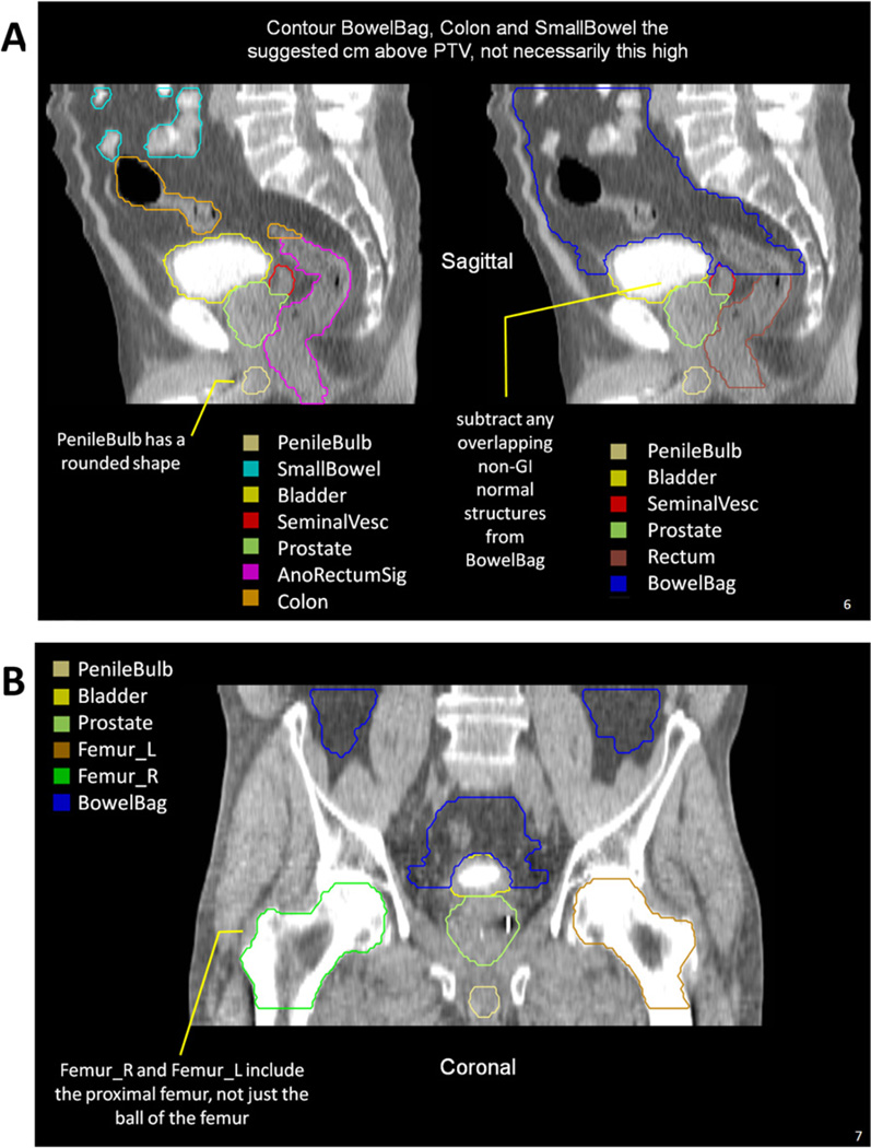
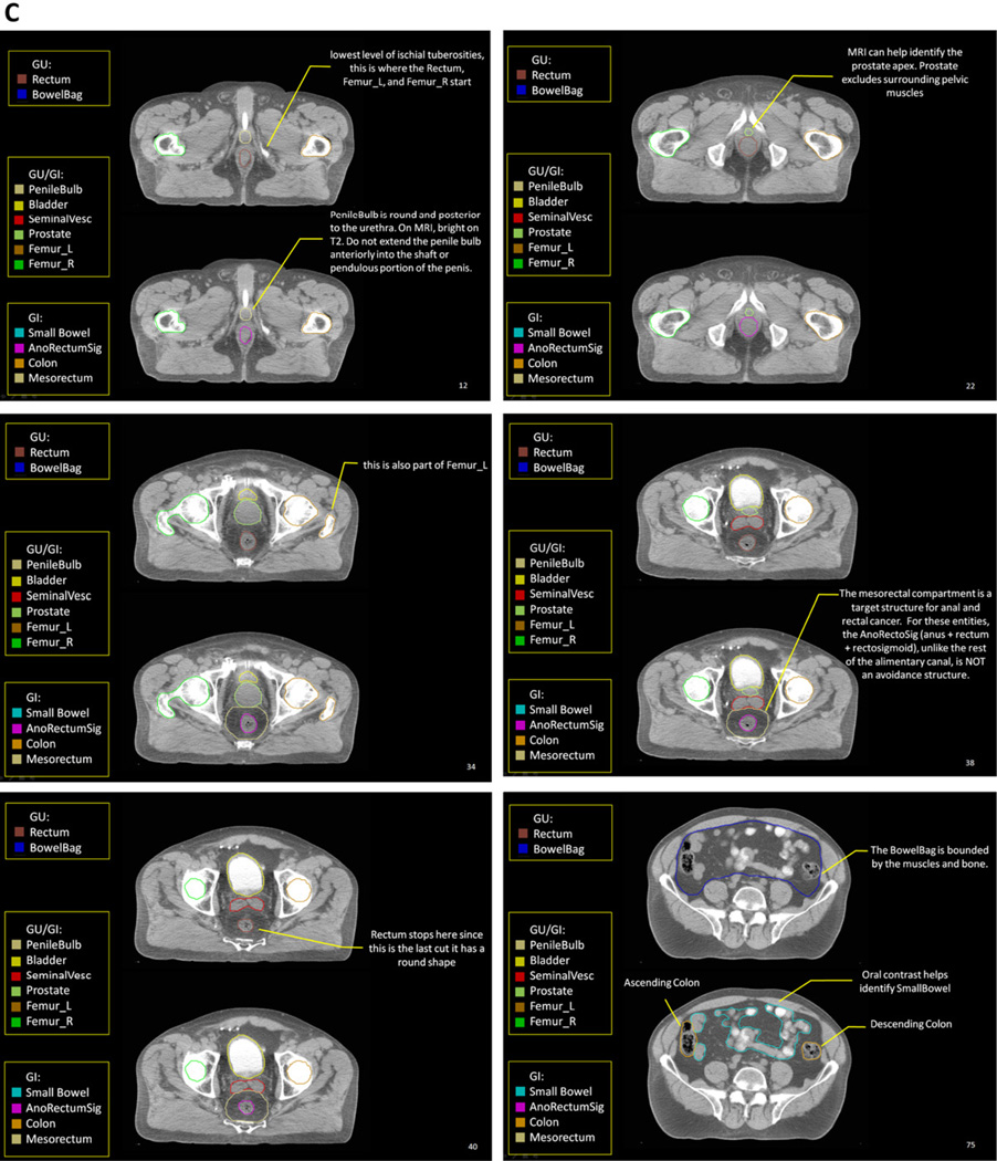
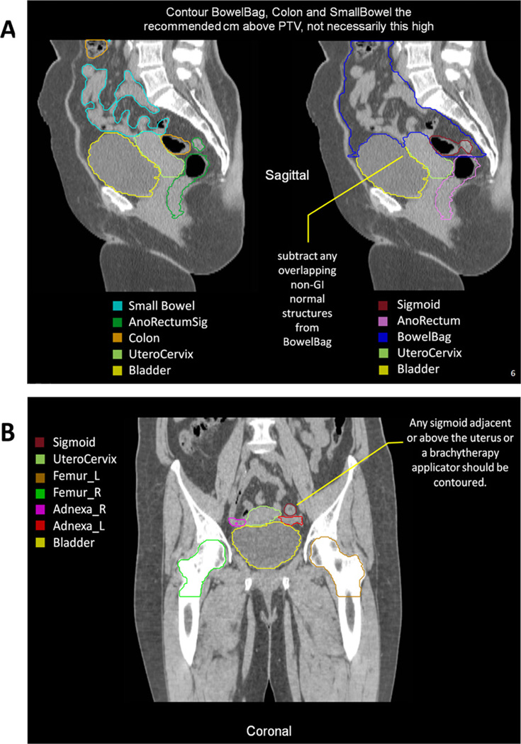
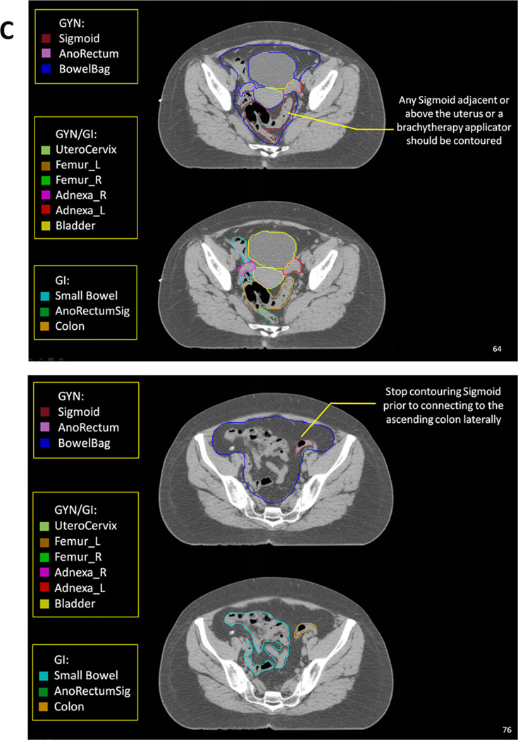
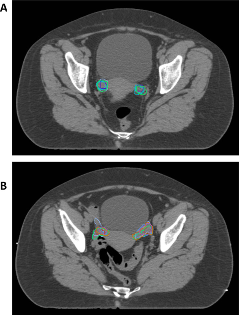
Methods and materials: One male pelvis computed tomography (CT) data set and one female pelvis CT data set were shared via the Image-Guided Therapy QA Center. A total of 16 radiation oncologists participated. The following organs at risk were contoured in both CT sets: anus, anorectum, rectum (gastrointestinal and genitourinary definitions), bowel NOS (not otherwise specified), small bowel, large bowel, and proximal femurs. The following were contoured in the male set only: bladder, prostate, seminal vesicles, and penile bulb. The following were contoured in the female set only: uterus, cervix, and ovaries. A computer program used the binomial distribution to generate 95% group consensus contours. These contours and definitions were then reviewed by the group and modified.
Results: The panel achieved consensus definitions for pelvic normal tissue contouring in RTOG trials with these standardized names: Rectum, AnoRectum, SmallBowel, Colon, BowelBag, Bladder, UteroCervix, Adnexa_R, Adnexa_L, Prostate, SeminalVesc, PenileBulb, Femur_R, and Femur_L. Two additional normal structures whose purpose is to serve as targets in anal and rectal cancer were defined: AnoRectumSig and Mesorectum. Detailed target volume contouring guidelines and images are discussed.
Conclusions: Consensus guidelines for pelvic normal tissue contouring were reached and are available as a CT image atlas on the RTOG Web site. This will allow uniformity in defining normal tissues for clinical trials delivering pelvic radiation and will facilitate future normal tissue complication research.
Copyright © 2012 Elsevier Inc. All rights reserved.
Conflict of interest statement
Conflict of interest: none.
Figures
References
-
- Deasy JO, Blanco AI, Clark VH. CERR: A computational environment for radiotherapy research. Med Phys. 2003;30:979–985. - PubMed
-
- McLaughlin PW, Evans C, Feng M, et al. Radiographic and anatomic basis for prostate contouring errors and methods to improve prostate contouring accuracy. Int J Radiat Oncol Biol Phys. 2010;76:369–378. - PubMed
Publication types
MeSH terms
Grants and funding
LinkOut - more resources
Full Text Sources
