. 2012 May 1;11(5):488-501.
doi: 10.1016/j.dnarep.2012.02.004. Epub 2012 Apr 6.

The conserved Cockayne syndrome B-piggyBac fusion protein (CSB-PGBD3) affects DNA repair and induces both interferon-like and innate antiviral responses in CSB-null cells

Affiliations

The conserved Cockayne syndrome B-piggyBac fusion protein (CSB-PGBD3) affects DNA repair and induces both interferon-like and innate antiviral responses in CSB-null cells

Arnold D Bailey et al. DNA Repair (Amst). .

Abstract

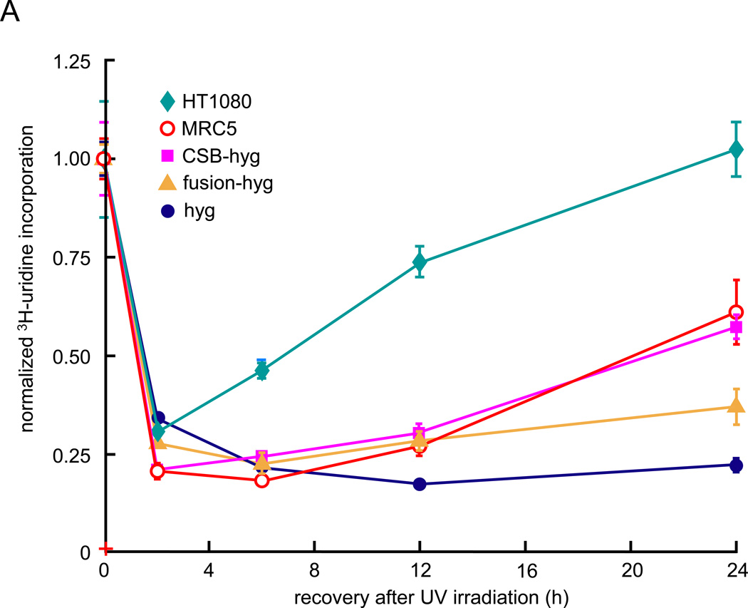
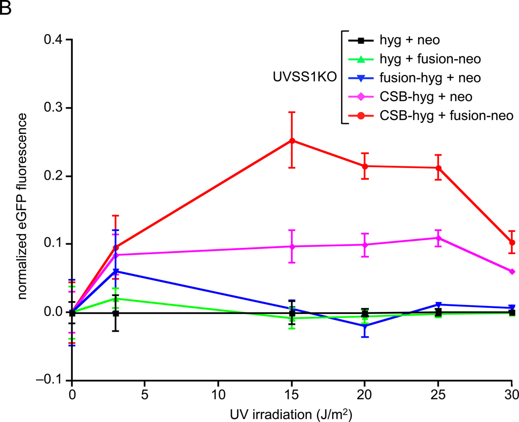
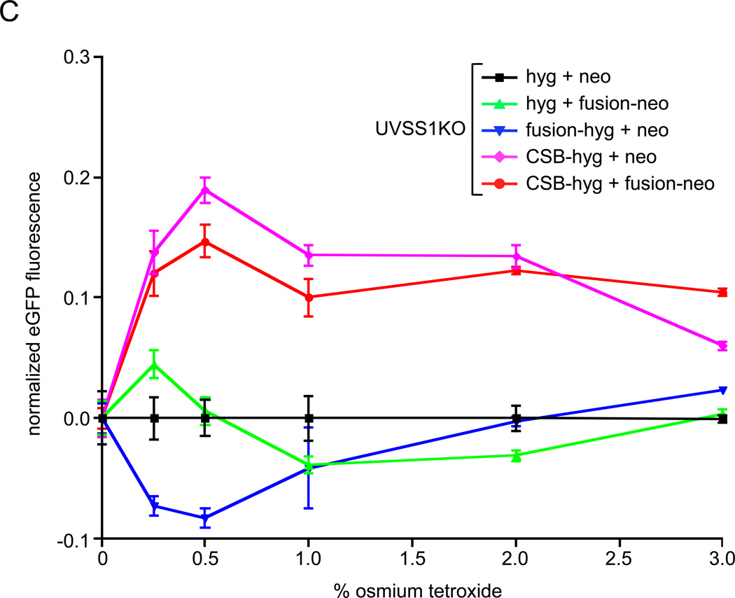
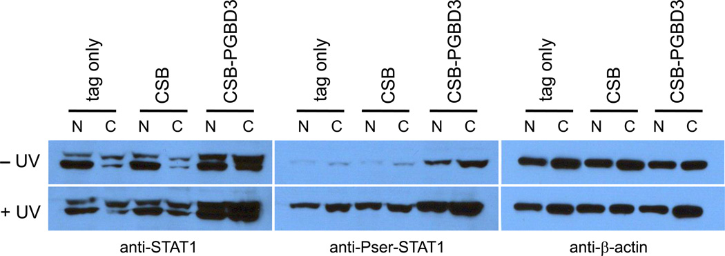
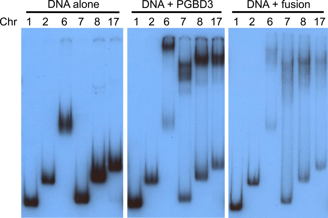
Cockayne syndrome is a segmental progeria most often caused by mutations in the CSB gene encoding a SWI/SNF-like ATPase required for transcription-coupled DNA repair (TCR). Over 43Mya before marmosets diverged from humans, a piggyBac3 (PGBD3) transposable element integrated into intron 5 of the CSB gene. As a result, primate CSB genes now generate both CSB protein and a conserved CSB-PGBD3 fusion protein in which the first 5 exons of CSB are alternatively spliced to the PGBD3 transposase. Using a host cell reactivation assay, we show that the fusion protein inhibits TCR of oxidative damage but facilitates TCR of UV damage. We also show by microarray analysis that expression of the fusion protein alone in CSB-null UV-sensitive syndrome (UVSS) cells induces an interferon-like response that resembles both the innate antiviral response and the prolonged interferon response normally maintained by unphosphorylated STAT1 (U-STAT1); moreover, as might be expected based on conservation of the fusion protein, this potentially cytotoxic interferon-like response is largely reversed by coexpression of functional CSB protein. Interestingly, expression of CSB and the CSB-PGBD3 fusion protein together, but neither alone, upregulates the insulin growth factor binding protein IGFBP5 and downregulates IGFBP7, suggesting that the fusion protein may also confer a metabolic advantage, perhaps in the presence of DNA damage. Finally, we show that the fusion protein binds in vitro to members of a dispersed family of 900 internally deleted piggyBac elements known as MER85s, providing a potential mechanism by which the fusion protein could exert widespread effects on gene expression. Our data suggest that the CSB-PGBD3 fusion protein is important in both health and disease, and could play a role in Cockayne syndrome.


Conflict of interest statement

Conflict of interest statement

The authors declare that there are no conflicts of interest.

Figures

Fig. 1
Fig. 1
piggyBacs are mobile DNA elements that survive as alternative 3' exons. (A) PGBD3 inserted into intron 5 of the primate CSB gene at least 43 Mya in the common ancestor of simian primates, with the result that the CSB gene now generates three proteins as shown in (B): full length CSB by default splicing of all 22 CSB exons, CSB-PGBD3 fusion protein by alternative splicing between CSB exon 5 and the PGBD3 alternative 3' terminal exon, and solitary PGBD3 driven by a cryptic promoter in CSB exon 5 [18]. The PGBD3 insertion generated a TTAA target site duplication. Immediately inside the subterminal inverted repeats of the mobile element, the transposase open reading frame (ORF) is flanked upstream by a 3' splice site (3' ss) and downstream by a polyadenylation site (pA). MER85 elements are nonautonomous internally-deleted PGBD3-derived elements that were last mobilized by a PGBD3-like transposase about 35 Mya [19]. CSB and PGBD3 sequences are indicated in blue and orange, respectively. The schematic not drawn to scale; the CSB gene spans 80 kb, the PGBD3 element 2.5 kb, and intact MER85s only 140 bp. (B) A comparison of the three proteins encoded by the CSB locus. The fusion protein joins the acidic 465 N-terminal residues of CSB exons 1–5, but none of the ATPase motifs (Roman numerals), to the 595 residue PGBD3 transposase.
Fig. 2
Fig. 2
Recovery of RNA synthesis (RRS) following UV damage, and host cell reactivation (HCR) assays for repair of oxidation- and UV-induced DNA damage. Assays were performed using CSB-null UVSS1KO-derived pools stably expressing CSB, CSB-PGBD3 fusion protein (two independent pools selected with either hygromycin or neomycin), both proteins, or tags only (Table 1). (A) RRS assays monitoring 3H-uridine incorporation after UV irradiation of growing cells with 10 J/m2/min. The SV40-immortalized normal lung cell line MRC5-SV and the the HT1080 fibrosarcoma cell linewere included as a controls [26]. (B) HCR assays for UV damage. The EGFP expression construct was irradiated with 1 J/m2/min UV from a germicidal lamp for 5–30 min before transfection. EGFP fluorescence was measured 48 h after transfection and normalized to cell number at each time point to correct for cell growth. (C) HCR assays for oxidative damage. The reporter EGFP expression construct was pretreated with the indicated concentrations of osmium tetroxide before transfection. Assays were performed in triplicate (panel A) or octuplicate (panels B and C); error bars smaller than the datapoint icons are not shown.
Fig. 2
Fig. 2
Recovery of RNA synthesis (RRS) following UV damage, and host cell reactivation (HCR) assays for repair of oxidation- and UV-induced DNA damage. Assays were performed using CSB-null UVSS1KO-derived pools stably expressing CSB, CSB-PGBD3 fusion protein (two independent pools selected with either hygromycin or neomycin), both proteins, or tags only (Table 1). (A) RRS assays monitoring 3H-uridine incorporation after UV irradiation of growing cells with 10 J/m2/min. The SV40-immortalized normal lung cell line MRC5-SV and the the HT1080 fibrosarcoma cell linewere included as a controls [26]. (B) HCR assays for UV damage. The EGFP expression construct was irradiated with 1 J/m2/min UV from a germicidal lamp for 5–30 min before transfection. EGFP fluorescence was measured 48 h after transfection and normalized to cell number at each time point to correct for cell growth. (C) HCR assays for oxidative damage. The reporter EGFP expression construct was pretreated with the indicated concentrations of osmium tetroxide before transfection. Assays were performed in triplicate (panel A) or octuplicate (panels B and C); error bars smaller than the datapoint icons are not shown.
Fig. 3
Fig. 3
Expression of the CSB-PGBD3 fusion protein in CSB-null UVSS1KO cells induces U-STAT1, which can be phosphorylated on serine 727 and further induced by UV irradiation. Using CSB-null UVSS1KO cells stably transfected with CSB, CSB-PGBD3 fusion protein, or tags alone (Table 1), nuclear and cytoplasmic fractions were resolved by SDS-PAGE, blotted, and probed with anti-STAT1 (left panel), anti-Pser-STAT1 (middle panel), and anti-β-actin antibodies as a loading control (right panel). The STAT1 locus generates two proteins (left panel): full length STAT1α (upper band) and C-terminally deleted STAT1β (lower band) lacking the Ser727 phosphorylation site. STAT1β has been referred to as a splice variant or proteolysis product [37]; however, mRNAs annotated on the UCSC Genome Browser (build hg18) indicate that STAT1β reflects alternative polyadenylation within intron 23. This results in use of a TAA terminator immediately following exon 23, and a protein that retains Tyr701 but lacks the Ser727 phosphorylation site.
Fig. 4
Fig. 4
The PGBD3 piggyBac transposase and the CSB-PGBD3 fusion protein bind to consensus MER85 elements in vitro. Electrophoretic mobility shift assays were performed [25] for binding of the recombinant PGBD3 and CSB-PGBD3 fusion proteins to 6 different genomic MER85 elements that closely match the 140 bp Repbase MER85 consensus. The multiple sequence alignment and genomic primers are shown in Fig. S2. Chromosome number is indicated above the lanes. MER85 elements from chromosomes 1 and 7 are MseI/MseI and AseI/HincII fragments, respectively, lacking flanking sequence; MER85s from chromosomes 2, 6, 8, and 17 are BamHI/EcoRI fragments that include the flanks. Although both proteins shift efficiently, the piggyBac transposase generates sharper bandshifts than the larger fusion protein on low percentage gels (6% 29:1 acrylamide:bisacrylamide for PGBD3 and 5% 80:1 acrylamide:bisacrylamide for CSB-PGBD3).

References

    1. Beerens N, Hoeijmakers JH, Kanaar R, Vermeulen W, Wyman C. The CSB protein actively wraps DNA. J Biol Chem. 2005;280:4722–4729. - PubMed
    1. Citterio E, Van Den Boom V, Schnitzler G, Kanaar R, Bonte E, Kingston RE, Hoeijmakers JH, Vermeulen W. ATP-dependent chromatin remodeling by the Cockayne syndrome B DNA repair-transcription-coupling factor. Mol Cell Biol. 2000;20:7643–7653. - PMC - PubMed
    1. Newman JC, Bailey AD, Weiner AM. Cockayne syndrome group B protein (CSB) plays a general role in chromatin maintenance and remodeling. Proc Natl Acad Sci U S A. 2006;103:9613–9618. - PMC - PubMed
    1. Nardo T, Oneda R, Spivak G, Vaz B, Mortier L, Thomas P, Orioli D, Laugel V, Stary A, Hanawalt PC, Sarasin A, Stefanini M. A UV-sensitive syndrome patient with a specific CSA mutation reveals separable roles for CSA in response to UV and oxidative DNA damage. Proc Natl Acad Sci U S A. 2009;106:6209–6214. - PMC - PubMed
    1. Groisman R, Kuraoka I, Chevallier O, Gaye N, Magnaldo T, Tanaka K, Kisselev AF, Harel-Bellan A, Nakatani Y. CSA-dependent degradation of CSB by the ubiquitin-proteasome pathway establishes a link between complementation factors of the Cockayne syndrome. Genes Dev. 2006;20:1429–1434. - PMC - PubMed

Publication types

MeSH terms