. 2012 May 8;22(9):743-52.
doi: 10.1016/j.cub.2012.02.066. Epub 2012 Apr 5.

Optogenetic analysis of a nociceptor neuron and network reveals ion channels acting downstream of primary sensors

Affiliations

Optogenetic analysis of a nociceptor neuron and network reveals ion channels acting downstream of primary sensors

Steven J Husson et al. Curr Biol. .

Abstract

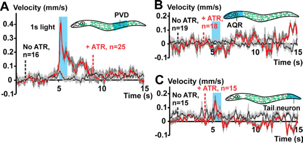
Background: Nociception generally evokes rapid withdrawal behavior in order to protect the tissue from harmful insults. Most nociceptive neurons responding to mechanical insults display highly branched dendrites, an anatomy shared by Caenorhabditis elegans FLP and PVD neurons, which mediate harsh touch responses. Although several primary molecular nociceptive sensors have been characterized, less is known about modulation and amplification of noxious signals within nociceptor neurons. First, we analyzed the FLP/PVD network by optogenetics and studied integration of signals from these cells in downstream interneurons. Second, we investigated which genes modulate PVD function, based on prior single-neuron mRNA profiling of PVD.

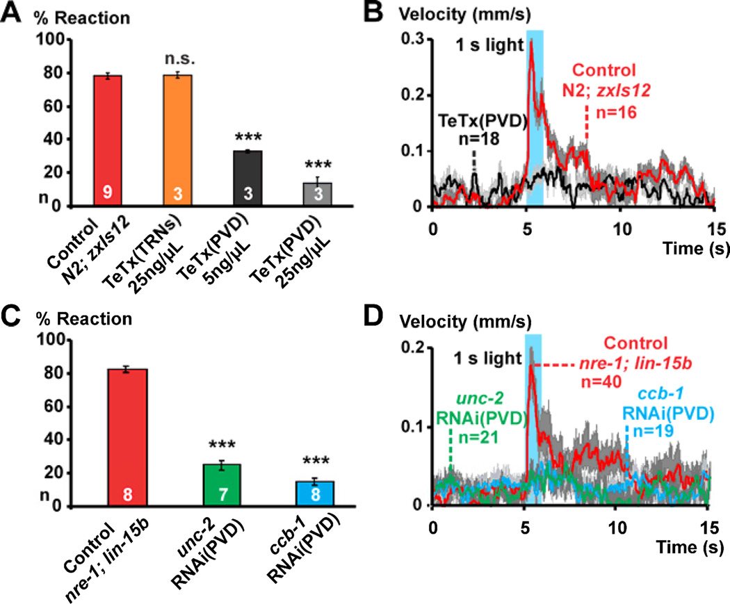
Results: Selectively photoactivating PVD, FLP, and downstream interneurons via Channelrhodopsin-2 (ChR2) enabled the functional dissection of this nociceptive network, without interfering signals by other mechanoreceptors. Forward or reverse escape behaviors were determined by PVD and FLP, via integration by command interneurons. To identify mediators of PVD function, acting downstream of primary nocisensor molecules, we knocked down PVD-specific transcripts by RNAi and quantified light-evoked PVD-dependent behavior. Cell-specific disruption of synaptobrevin or voltage-gated Ca(2+) channels (VGCCs) showed that PVD signals chemically to command interneurons. Knocking down the DEG/ENaC channel ASIC-1 and the TRPM channel GTL-1 indicated that ASIC-1 may extend PVD's dynamic range and that GTL-1 may amplify its signals. These channels act cell autonomously in PVD, downstream of primary mechanosensory molecules.

Conclusions: Our work implicates TRPM channels in modifying excitability of and DEG/ENaCs in potentiating signal output from a mechano-nociceptor neuron. ASIC-1 and GTL-1 homologs, if functionally conserved, may denote valid targets for novel analgesics.


Figures

Figure 1
Figure 1. Photostimulation of PVD results in forward escape behavior
(A) Confocal stacks of an adult animal carrying transgene zxIs12[pF49H12.4::ChR2::mCherry; pF49H12.4::GFP], expressing GFP and ChR2::mCherry in PVD (cell body and branched arbors), in addition to neuron AQR and one unidentified tail neuron. (B) lite-1(ce314); zxIs12 animals were challenged with blue light pulses of 0.2, 1 and 5s (indicated by blue shading). Resulting escape behavior was quantified using a video analysis tool [43]; mean speeds and SEMs are shown. Animals cultivated without the ChR2 cofactor all-trans-retinal (ATR) showed no responses. (C) Fractions of animals reacting to increasing light intensities are shown for different developmental stages (larval stages L2-L4, and adult; n>30 each).
Figure 2
Figure 2. Selective illumination of PVD cell body and of other cells expressing ChR2
(A–C) Selective illumination of predefined body segments of freely moving wild type (N2); zxIs12 animals was used as indicated in pictograms, to restrict light to the cell bodies of PVD or other neurons expressing ChR2. Mean velocity traces and SEMs are shown.
Figure 3
Figure 3. Both mechanical and optical nociceptive harsh touch stimuli elicit less habituation than gentle touch stimuli
(A) Fractions of wild type (N2); zxIs12 animals (black) and animals expressing ChR2 in the TRNs (Pmec-4::ChR2, gray), were assessed for escape responses to 20 successive light pulses of 0.5s with 9.5s interstimulus interval (n≥25). (B) Wild type animals were challenged with harsh or gentle touch every 10s, and fractions of animals reacting were quantified (n=20). Regression analysis shows different slopes for the fitted lines in both optical and mechanical touch experiments (*p<0.05).
Figure 4
Figure 4. Functional analysis of the PVD and FLP harsh touch nociceptor network
(A) PVD was co-stimulated with anterior TRNs ALM and AVM by selective illumination using variable light intensities as represented by the blue arrows below the graph (see also Figure S3A–I). Fractions of tested animals responding with forward or backward escape are shown. (B) Simplified neuronal wiring diagram based on Figure S1, displaying synaptic connections from PVD and FLP sensory neurons (triangles) and their integration into the command neuron (hexagons) and motor neuron circuits (circles) [12]. Numbers of synapses, which could be excitatory, inhibitory, or both, are shown. (C) Ca2+-imaging in PVC (using RCaMP), before and after a 1s photostimulus to PVD (blue bar, lower panel). Fluorescent signals (mean ΔF/F±SEM) in PVC are compared to red fluorescence of ChR2:mCherry expressed from the pF49H12.4 promoter in a tail neuron next to PVC (ROIs indicated in upper panel; see also Movie S4). (D) Fractions of animals that moved forward, backward, or did not respond upon photoactivation of PVD (zxIs12) in the wild type (N2) or in deg-1(d) mutants (PVC forward command neurons degenerated). Similarly, behaviors evoked by anterior or posterior mechanical harsh touch are indicated for deg-1(d), mec-4(d) (lacking TRNs) and wild type (N2) animals; n≥120; ***p<0.0001, **p<0.005, n.s. not significant (Chi2-tests). (E) Selective illumination and video tracking was used to record behaviors induced by PVD photoactivation in wild type (N2); zxIs12 animals or deg-1(d); zxIs12 animals. (F) Illuminating the head of animals expressing Pegl-46::ChR2::YFP, photoactivating FLP neurons, evokes backward movement. In E, F, mean velocity±SEM are shown.
Figure 5
Figure 5. Optogenetics-assisted functional RNAi screen of PVD-enriched genes
(A) We previously isolated mRNAs from PVD neurons, using the mRNA tagging method as reported in Smith et al. [19]. Epitope (FLAG) tagged poly-A binding protein (PAB-1) was expressed in PVD (and OLL) neurons, using the ser-2prom3 promoter. PAB-1-mRNA was immunoprecipitated and compared to mRNA isolated from all cells by microarray profiling. Candidate genes were knocked down by RNAi and photoevoked PVD-dependent escape behaviors assessed. (B) For each knockdown line (or genomic mutant), 25–30 young adults were assessed for blue light-evoked escape behavior and fractions of animals reacting were compared to negative controls (animals fed with the empty L4440 RNAi vector). Each trial was repeated several times, and normalized to the respective negative control done on the same day. Bars are color-coded according to x-fold mRNA enrichment in PVD (and OLL neurons, compared to all other cells, see Table S1). ANOVA with Tukey’s post-hoc test; ***p<0.0001, **p<0.005, *p<0.05.
Figure 6
Figure 6. Inhibition of synaptic signaling and cell-specific knockdown of unc-2 and ccb-1 impair PVD function
(A, B) Animals with impaired synaptic signaling were generated by injecting wild type (N2); zxIs12 animals (control) with the coding sequence of Tetanus toxin light chain using the mec-4 promoter to achieve expression in TRNs (TeTx(TRNs), 25ng/µL), or using the PVD-specific promoter F49H12.4 at two different concentrations (TeTx(PVD) 5 and 25ng/µL). (A) Fractions of animals reacting for each strain upon 0.2mW/mm2 blue light pulses. (B) A 1s blue light pulse was applied to the nematode segment with PVD cell bodies and resulting locomotion behavior was quantified. Mean velocities±SEM (gray) are shown. (C–D) Cell-specific knockdown of unc-2 or ccb-1 was achieved as described [26] (see Figure S5A) (C) Fractions of animals reacting and (D) velocity diagrams are shown. Paired t-test; ***p<0.0001.
Figure 7
Figure 7. ASIC-1 and GTL-1 accentuate and amplify ChR2-evoked signals in PVD
(A) Different light intensities were used to stimulate PVD by whole body illumination, and the fraction of animals reacting was quantified (25–30 animals per trial) for each genotype or cell-specific knockdown (n=8 for wild type (N2); n=4 each for asic-1 RNAi(PVD) and gtl-1 RNAi(PVD)). Error bars indicate SEM. Boltzmann sigmoidals were fitted according to [%reacting worms = %max / (1+exp((I50-I)/Slope))], where I is light intensity, %max is the maximal fraction of worms reacting and I50 indicates the light intensity at %max/2. Statistical differences for the three free parameters, %max, I50 and slope (indicated in the table) were determined by regression analysis; *p<0.05, **p<0.005, n.s. not significant. (B) The onset of escape behavior for a 1s illumination of the nematode segment with PVD cell bodies is shown from the time of illumination (indicated as 0s) to 0.5s after illumination. Mean velocity traces for N2, asic-1 RNAi(PVD), gtl-1 RNAi(PVD), asic-1(ok415) and gtl-1(ok375) can be fitted by Boltzmann sigmoidals until the maximal velocity is reached according to [velocity = max velocity / (1+exp((T50-T)/Slope))]. (C) PVD cell bodies of ≥20 animals were photoactivated for 1s by selective illumination for different light intensities; see Figure S6 for all velocity traces. Mean maximal velocities±SEM for all light intensities are plotted for each genotype; t-test; *p<0.05, **p<0.005. (D) Fractions of animals responding are displayed for each light intensity, corresponding Boltzmann sigmoidal fits and free parameters are indicated; *p<0.05.

References

    1. Basbaum AI, Bautista DM, Scherrer G, Julius D. Cellular and molecular mechanisms of pain. Cell. 2009;139:267–284. - PMC - PubMed
    1. Kang L, Gao J, Schafer WR, Xie Z, Xu XZ. C. elegans TRP family protein TRP-4 is a pore-forming subunit of a native mechanotransduction channel. Neuron. 2010;67:381–391. - PMC - PubMed
    1. Li W, Kang L, Piggott BJ, Feng Z, Xu XZ. The neural circuits and sensory channels mediating harsh touch sensation in Caenorhabditis elegans. Nat Commun. 2011;2:315. - PMC - PubMed
    1. O'Hagan R, Chalfie M, Goodman MB. The MEC-4 DEG/ENaC channel of Caenorhabditis elegans touch receptor neurons transduces mechanical signals. Nat. Neurosci. 2005;8:43–50. - PubMed
    1. Tobin DM, Bargmann CI. Invertebrate nociception: behaviors, neurons and molecules. J. Neurobiol. 2004;61:161–174. - PubMed

Publication types