. 2012 May 8;22(9):753-62.
doi: 10.1016/j.cub.2012.02.069. Epub 2012 Apr 5.

EB1-recruited microtubule +TIP complexes coordinate protrusion dynamics during 3D epithelial remodeling

Affiliations

EB1-recruited microtubule +TIP complexes coordinate protrusion dynamics during 3D epithelial remodeling

Sarah Gierke et al. Curr Biol. .

Abstract

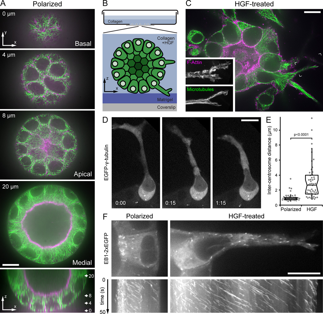
Background: Epithelial remodeling, in which apical-basal polarized cells switch to a migratory phenotype, plays a central role in development and disease of multicellular organisms. Although dynamic microtubules (MTs) are required for directed migration on flat surfaces, how MT dynamics are controlled or contribute to epithelial remodeling in a more physiological three-dimensional (3D) environment is not understood. We use confocal live-cell imaging to analyze MT function and dynamics during 3D epithelial morphogenesis and remodeling of polarized Madin-Darby canine kidney epithelial cells that undergo partial epithelial-to-mesenchymal transition in response to hepatocyte growth factor (HGF).

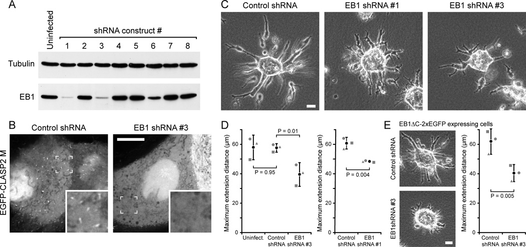
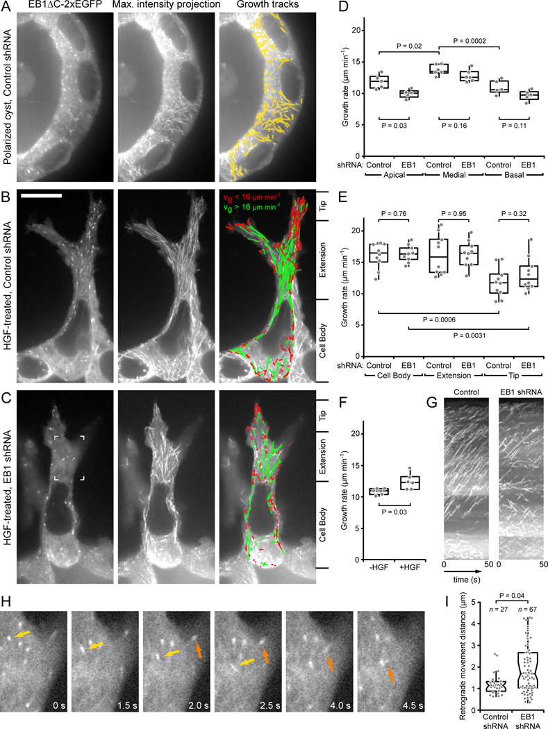
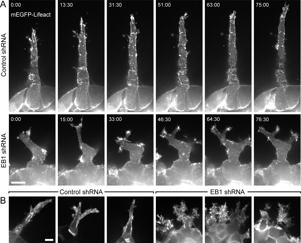
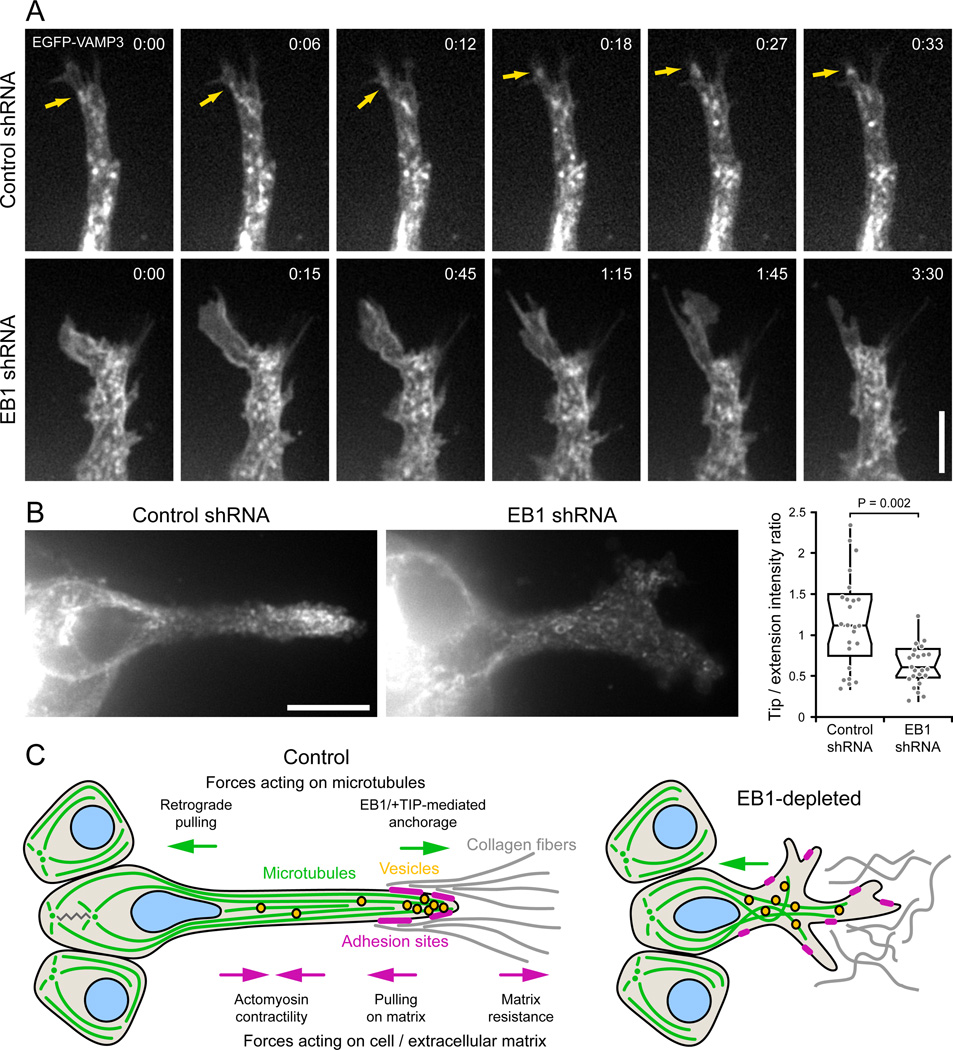
Results: We find that HGF treatment increases MT growth rate before morphological changes are evident and that large numbers of MTs grow into HGF-induced cell extensions independent of centrosome reorientation. Using lentivirus-mediated small hairpin RNA, we demonstrate that EB1, an adaptor protein that mediates recruitment of numerous other +TIP proteins to growing MT plus ends, is required for this HGF-induced MT reorganization. We further show that protrusion and adhesion dynamics are disorganized and that vesicular trafficking to the tip of HGF-induced cell extensions is disrupted in EB1-depleted cells.

Conclusions: We conclude that EB1-mediated interactions with growing MTs are important to coordinate cell shape changes and directed migration into the surrounding extracellular matrix during epithelial remodeling in a physiological 3D environment. In contrast, EB1 is not required for the establishment or maintenance of apical-basal cell polarity, suggesting different functions of +TIPs and MTs in different types of cell polarity.


Figures

Figure 1
Figure 1. Microtubule cytoskeleton reorganization during HGF-induced epithelial remodeling
(A) Spinning disk confocal microscopy optical sections of a polarized MDCK cyst stained for F-actin (magenta) and MTs (green). Distance from the bottom of the cyst is indicated. The bottom panel is a z-axis reconstruction, and the approximate location of the optical sections shown is indicated. (B) Diagram of the 3D MDCK epithelial culture system adapted for high-resolution imaging. (C) Single optical section of F-actin (magenta) and MTs (green) 24 hours after HGF addition. Insets show a maximum intensity projection of the indicated area at higher magnification to illustrate the dense MT array extending to the extension tip. (D) EGFP-γ-tubulin time-lapse sequence in an HGF-induced extension showing centrosome separation and dynamics. Centrosomes remain close in the neighboring polarized cell that has not formed an extension. Elapsed time is indicated in hours:minutes. (E) Distance between centrosomes in untreated polarized cells and cells with HGF-induced extensions. Gray symbols are measurements from individual cells (n = 50). (F) Images from medial optical sections of EB1-2xEGFP time-lapse sequences recorded at 2 frames s−1 with the apical surface oriented to the left. The bottom panels show kymographs perpendicular to the apical-basal cell axis. Oblique lines represent MT ends growing toward the basal surface or into the HGF-induced extension, respectively. Scale bars, 10 µm. See also Fig. S1 and Movie 1.
Figure 2
Figure 2. EB1 is required for HGF-induced epithelial remodeling
(A) Immunoblot of lysates from cells expressing different shRNA constructs after 7 days of puromycin selection. Tubulin was used as loading control. (B) MDCK cells stably expressing control or EB1 shRNA#3, transiently transfected with an EGFP-tagged, non-phosphorylatable construct encoding the CLASP2 EB1-binding domain (CLASP2 M) [8]. Insets show indicated region at higher magnification. Scale bar, 10 µm. (C) Phase contrast images of control and EB1-depleted HGF-treated MDCK cysts. Scale bar, 20 µm. (D) Quantification of HGF-induced cell extension into the surrounding collagen matrix. Graphs shows the average length of the three longest extensions measured from extension tip to cyst edge. Gray symbols are the average of 80–100 cysts from 3 separate experiments. Black circle represents the average of these three experiments. Error bars indicate 95% confidence interval. (E) Phase contrast images of EB1ΔC-2xEGFP-expressing control and EB1-depleted HGF-treated MDCK cysts, and quantification of HGF-induced cell extensions as in (D). Scale bar, 20 µm. See also Fig. S2 and Movie 2.
Figure 3
Figure 3. Analysis of microtubule dynamics in 3D epithelial structures
(A–C) Representative images of EB1ΔC-2xEGFP tracking in (A) a polarized, control shRNA cyst, and HGF-induced extensions of (B) control and (C) EB1-depleted cysts. Left panels are single images of the time-lapse sequence, middle panels show maximum intensity projections over the entire sequence (100 frames recorded at 2 frames s−1), and right panels show computer-generated tracks overlaid on the maximum intensity projection indicating tracking quality. Only tracks with a lifetime of >2.5 s are shown. Tracks in (B) and (C) are color-coded for growth rate. (D) MT growth rates in apical, medial, and basal optical sections in control or EB1-depleted polarized cysts. (E) MT growth rates in the cell body, extension and extension tip in control or EB1-depleted HGF-induced extensions. Division of cells into these three regions is indicated on the right of (B) and (C). (F) MT growth rates in basal optical sections in polarized cysts in Matrigel expressing EB1-2xEGFP before and 5 hrs after HGF addition. (D–F) Gray symbols represent the average growth rate of all tracks per cell and indicated condition. P-values of relevant comparisons are indicated on the graphs. (G) Kymographs of the cells shown in (B) and (C). (H) EB1ΔC-2xEGFP time-lapse sequence in an EB1-depleted HGF-induced extension. The region shown is indicated in (C). Two growing MT ends that display extensive retrograde movements are highlighted by arrows. Elapsed time is in seconds. (I) Number and distance of all retrograde MT movements in ten control and EB1-depleted HGF-induced extensions. Each gray symbol represents a retrograde movement event. Scale bar, 10 µm. See also Fig. S3, Movies 3, 4 and 5.
Figure 4
Figure 4. Uncoordinated protrusion dynamics in EB1-depleted cells
(A) mEGFP-Lifeact time-lapse sequences of HGF-induced extensions in control and EB1-depleted cysts. Each image is a maximum intensity projection of three optical sections (2 µm between sections) to compensate for cell movement out of the plane of focus. Elapsed time is in minutes:seconds. (B) Maximum intensity projection over two hour time-lapse sequences, which highlights increased and uncoordinated actin dynamics in EB1-depleted cells. Scale bars, 10 µm. See also Movie 6.
Figure 5
Figure 5. EB1 is required for productive interactions with the extracellular matrix
(A) Paxillin-EGFP time-lapse sequences of HGF-induced extensions in control and EB1-depleted cysts. Each image is a maximum intensity projection of three optical sections (1 µm between sections). Insets show higher magnification of indicated areas. (B) Axial ratio, defined as long divided by short axis, of focal adhesions in control and EB1-depleted extensions. The three longest adhesions were measured for each extension and each circle represents one adhesion (n = 60 adhesions in 20 cells). (C) DIC time-lapse sequences of HGF-induced extensions protruding into the surrounding fibrillar collagen matrix. Arrows indicate movement of fiduciary marks in the collagen between first and last frame shown. Elapsed time is indicated in minutes:seconds in (A) and (C). (D) pMLC immunofluorescence in HGF-induced extensions. Each image is a maximum intensity projection of three optical sections (1 µm between sections). Arrows indicate pMLC staining along cortical actin cables in control cells. (E) Ratio of pMLC fluorescence intensity along cell extension edge and cytoplasm. (n = 20 cell extensions). See also Movies 7 and 8. Scale bars, 10 µm.
Figure 6
Figure 6. EB1 depletion disrupts VAMP3-positive vesicle trafficking to the extension tip
(A) EGFP-VAMP3 time-lapse sequences of control and EB1-depleted HGF-induced extensions. Faster time scale in control cell demonstrates the rapid movement of vesicles into the extension tip, while the membrane protrusion in the EB1-depleted cell remains vacant over a much longer time period. Arrows indicate vesicle movement into the extension tip. Elapsed time is indicated in minutes:seconds. Scale bar, 5 µm. (B) VAMP3-positive vesicles accumulate near the extension tip in control cells. The graph shows a quantification of the ratio of EGFP-VAMP3 fluorescence intensity near the extension tip and in the middle of the cell extension (n = 25 cells). Scale bar, 10 µm. (C) Model of EB1 contribution to cell migration in a 3D environment. EB1-recruited +TIP complexes anchor and stabilize MTs along the HGF-induced cell extension, providing mechanical resistance to contractile forces in the cell body and establishing transport tracks toward the extension tip.

References

    1. Bryant DM, Mostov KE. From cells to organs: building polarized tissue. Nat. Rev. Mol. Cell Biol. 2008;9:887–901. - PMC - PubMed
    1. Affolter M, Zeller R, Caussinus E. Tissue remodelling through branching morphogenesis. Nat. Rev. Mol. Cell Biol. 2009;10:831–842. - PubMed
    1. Kaverina I, Straube A. Regulation of cell migration by dynamic microtubules. Semin. Cell Dev. Biol. 2011;22:968–974. - PMC - PubMed
    1. Wittmann T, Waterman-Storer CM. Cell motility: can Rho GTPases and microtubules point the way? J. Cell Sci. 2001;114:3795–3803. - PubMed
    1. Myers KA, Applegate KT, Danuser G, Fischer RS, Waterman CM. Distinct ECM mechanosensing pathways regulate microtubule dynamics to control endothelial cell branching morphogenesis. J. Cell Biol. 2011;192:321–334. - PMC - PubMed

Publication types

MeSH terms

Substances