. 2012 Jul;184(1):39-43.
doi: 10.1016/j.molbiopara.2012.03.006. Epub 2012 Mar 30.

Protein palmitoylation inhibition by 2-bromopalmitate alters gliding, host cell invasion and parasite morphology in Toxoplasma gondii

Affiliations

Protein palmitoylation inhibition by 2-bromopalmitate alters gliding, host cell invasion and parasite morphology in Toxoplasma gondii

A M Alonso et al. Mol Biochem Parasitol. 2012 Jul.

Abstract

Protein palmitoylation is the reversible covalent attachment of palmitic acid onto proteins. This post-translational modification has been shown to play a part in diverse processes such as signal transduction, cellular localization and regulation of protein activity. Although many aspects of protein palmitoylation have been identified in mammalian and yeast cells, little is known of this modification in Toxoplasma gondii. In order to determine the functional role of protein palmitoylation in T. gondii, tachyzoites were treated with the palmitoylation inhibitor 2-bromopalmitate (2-BP). Parasites treated with 2-BP displayed a significant increase in non-circular trails which were longer than those trails left by non-treated parasites. Furthermore, 2-BP treatment reduced the invasion process to the host cells. Long-term treatment of intracellular tachyzoites resulted in major changes in parasite morphology and shape in a dose-dependent manner. These results suggest that palmitoylation could be modifying proteins that are key players in gliding, invasion and cytoskeletal proteins in T. gondii.


Figures

Fig. 1
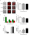
Fig. 1. Analysis of protein depalmitoylation on extracellular tachyzoites
(A) Parasites (2×10⁶) were treated either with DMSO (control), 50 μM or 100 μM of 2-BP for 2 hours and resuspended in 150 μl DMEM+P+S and subjected to immunofluorescence studies using anti-SAG1 antibody. All observations were performed on a Nikon E600 epifluorescence microscope. Adobe Photoshop (Adobe Systems) was used for image processing. PC: phase contrast. A: 100X. (B) After 2 hours of 2-BP treatment, T. gondii’s viability was assessed by the XTT method, following the manufacturer’s instructions. The percentage of optical density was calculated in respect to control. Three independent experiments in quintuplicate were performed and analyzed. White bar: control (DMSO treated); grey bar: 50 μM 2-BP; black bar: 100 μM 2-BP. (C) Parasites were treated for 2 hours with 2-BP, washed and allowed to invade fibroblast monolayers for 1 hour at 37°C. They were then fixed and incubated with rabbit anti-SAG1 as extracellular parasite labeling antibody. After that, the samples were permeabilized by including 0.1% (v/v) TX-100 in the PBS–BSA block and incubated with murine anti-IMC1 antibody. Secondary antibodies used were Alexafluor goat anti-mouse 488 and Alexafluor goat anti-rabbit 594 (Invitrogen). For each treatment, at least 250 total parasites were counted from nine random fields per sample, and values are presented as internal (green) or external (red) parasites per field. Data are mean values +/- SEM (error bars) for three independent experiments performed in duplicates. Different letters indicate significant differences with a p<0.05. Intracellular and extracellular parasites were analyzed separately using one-way ANOVA followed by Tukey’s multiple comparison tests. Prism GraphPad software was used for statistical analysis. (D) Trails left for each of the concentration tested after 2 hour 2-BP treatment were analyzed by indirect immunofluorescence using anti-SAG1 antibody and performed on poly-l-lysine coated slides. Percentage of the parasites associated to trails in regards to the total of parasites did not differ significantly at the concentration tested. Three independent experiments in duplicate were performed and 100 fields were analyzed each time. (E) From all the parasites associated to trails, the percentage of circular versus non-circular trails was calculated. Significant differences could be observed at 50 μM 2-BP. White bar: control (DMSO treated); grey bar: 50 μM 2-BP; black bar: 100 μM 2-BP. Different letters indicate significant differences with a p< 0.05. Three independent experiments in duplicate were performed and an average of 100 trails was analyzed. Non-circular trails (10 for each concentration) were chosen at random and analyzed (see Fig. S1). (F) Quantification of trail-lengths left after treatment with 2-BP. Significant differences could be observed in a dose-dependent manner. White bar: control (DMSO treated); grey bar: 50 μM 2-BP; black bar: 100 μM 2-BP. Different letters indicate significant differences with a p< 0.05. ANOVA analysis, followed by Tukey’s multiple comparison tests was performed with the data obtained at least in three independent experiments with duplicates in each.
Fig. 2
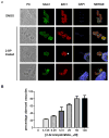
Fig. 2. Analysis of protein depalmitoylation on intracellular tachyzoites
(A) Untreated tachyzoites were incubated with a monolayer of HFF cells. After 1 hour at 37°C in 5% CO2, the wells were washed three times with PBS and fresh media containing 0, 3.125, 6.25, 12.5, 25, 50 and 100 μM of 2-BP in DMSO was added to the cells. Sixteen hours post-infection cells were fixed and permeabilized. Dividing tachyzoites were visualized as budding cells labeled with anti-IMC1 antibody, whereas nuclei were visualized with DAPI. Morphology could be observed by anti-SAG1 antibody. PC: phase contrast. A100×. (B) Percentage of intracellular parasites with aberrant morphology was plotted. This figure is representative of six independent experiments performed in duplicates where at least 200 vacuoles were counted. All the experiments presented similar results (see Fig. S2 to observe aberrant vacuoles at each concentration tested).

References

    1. Linder ME, Deschenes RJ. New insights into the mechanisms of protein palmitoylation. Biochemistry. 2003;42:4311–20. - PubMed
    1. Edidin M. The state of lipid rafts: from model membranes to cells. Annu Rev Biophys Biomol Struct. 2003;32:257–83. - PubMed
    1. Corvi MM, Soltys CL, Berthiaume LG. Regulation of mitochondrial carbamoyl-phosphate synthetase 1 activity by active site fatty acylation. J Biol Chem. 2001;276:45704–12. - PubMed
    1. Casey PJ. Protein lipidation in cell signaling. Science. 1995;268:221–5. - PubMed
    1. Apolloni A, Prior IA, Lindsay M, Parton RG, Hancock JF. H-ras but not K-ras traffics to the plasma membrane through the exocytic pathway. Mol Cell Biol. 2000;20:2475–87. - PMC - PubMed

Publication types

MeSH terms

LinkOut - more resources