

doi: 10.1038/nsmb.2270.
Crystal structure of a group II intron in the pre-catalytic state
Affiliations
- PMID: 22484319
- PMCID: PMC3670821
- DOI: 10.1038/nsmb.2270
Item in Clipboard
Crystal structure of a group II intron in the pre-catalytic state
Nat Struct Mol Biol.
.
Abstract
Group II introns are self-splicing catalytic RNAs that are thought to be ancestral to the spliceosome. Here we report the 3.65-Å crystal structure of the group II intron from Oceanobacillus iheyensis in the pre-catalytic state. The structure reveals the conformation of the 5' splice site in the catalytic core and represents the first structure of an intron prior to the first step of splicing.
Figures
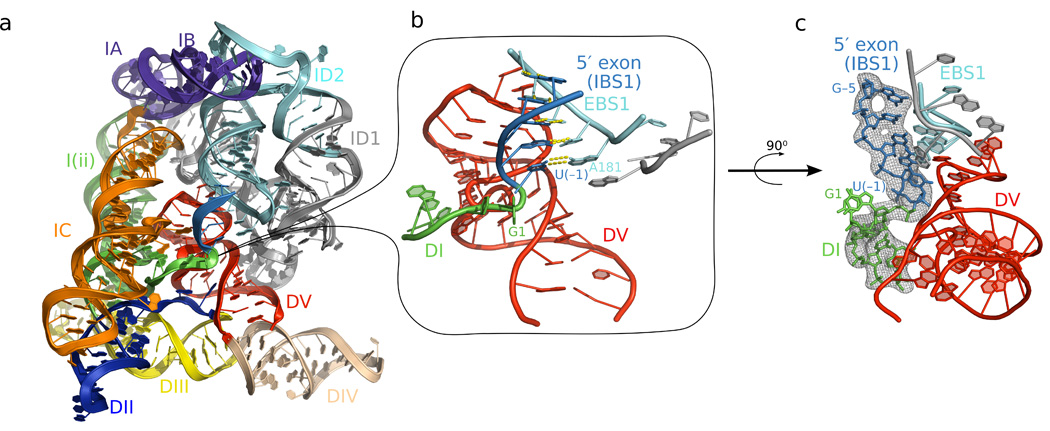
The pre-catalytic structure of the group II intron. a) The structure of the
pre-catalytic state reveals the position of the 5′ splice site (junction between green and
blue) in the center of the molecule. b) Close-up view of the 5′ splice site reveals a sharp
kink in the backbone, which is positioned near the bulge and catalytic triad of domain V (red). c)
Fo-Fc density for the 5′ splice site contoured at 3σ. The
Fo-Fc density map was calculated using a model deleted for intron nucleotides
1–5 and the 5′ exon in order to avoid model bias.

Theoretical model for the complete group II intron splicing pathway. a) The 5′
splice site is kinked immediately before the onset of catalysis. The kink is positioned in close
proximity to the catalytic metal ions (M1 and M2). A ribose 2′-OH group or water molecule
(not depicted) is activated for nucleophilic attack and cleaves the splice site. Residue 288 is not
shown because there is no electron density for the base of this nucleotide in the pre-catalytic
structure. b) The 3′ splice site is positioned in the active site through its interaction
with EBS3 and the γ nucleotide. These tertiary interactions cause the 3′ splice site
to also adopt a sharp kink which presents the scissile phosphate to the active site metal ions. The
3′-OH (shown in stick format) of the 5′ exon is in a position to coordinate to M1 as
well as to the 3′ splice site (coordination indicated by black lines). G1 and U2 are not
shown due to the fact that these nucleotides must depart before the 3′ splice site enters
the active site. c) The 3′ splice site is cleaved, the exons ligated, and the product adopts
a “relaxed” conformation prior to release by the intron. Abbreviation: J2/3 -
junction sequence between domains II and III.
References
-
- Sharp PA. Five easy pieces. Science. 1991;254:663. - PubMed
-
- Martin W, Koonin EV. Introns and the origin of nucleus-cytosol compartmentalization. Nature. 2006;440:41–45. - PubMed
-
- Robart AR, Zimmerly S. Group II intron retroelements: function and diversity. Cytogenet Genome Res. 2005;110:589–597. - PubMed
-
- Jacquier A, Michel F. Multiple exon-binding sites in class II self-splicing introns. Cell. 1987;50:17–29. - PubMed
Publication types
MeSH terms
Substances
Associated data
- Actions
Grants and funding
LinkOut - more resources
Full Text Sources
Other Literature Sources
