Review
. 2012 Sep;23(7):729-37.
doi: 10.1016/j.semcdb.2012.03.014. Epub 2012 Mar 30.

Intestinal redox biology and oxidative stress

Affiliations
Review

Intestinal redox biology and oxidative stress

Magdalena L Circu et al. Semin Cell Dev Biol. 2012 Sep.

Abstract

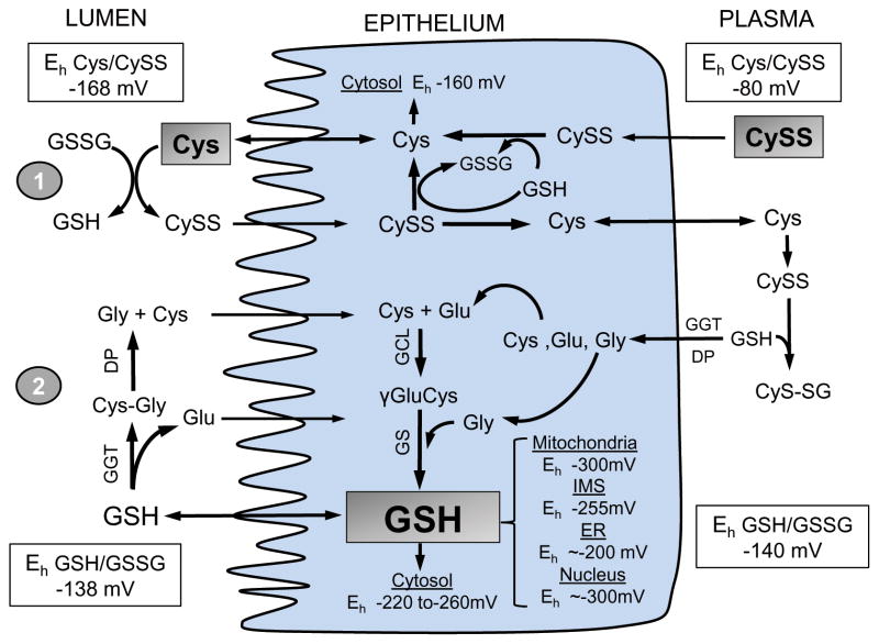
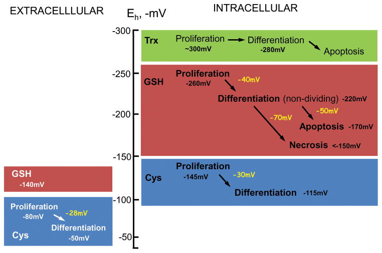
The intestinal epithelium sits at the interface between an organism and its luminal environment, and as such is prone to oxidative damage induced by luminal oxidants. Mucosal integrity is maintained by the luminal redox status of the glutathione/glutathione disulfide (GSH/GSSG) and cysteine/cystine (Cys/CySS) couples which also support luminal nutrient absorption, mucus fluidity, and a diverse microbiota. The epithelial layer is uniquely organized for rapid self-renewal that is achieved by the well-regulated processes of crypt stem cell proliferation and crypt-to-villus cell differentiation. The GSH/GSSG and Cys/CySS redox couples, known to modulate intestinal cell transition through proliferation, differentiation or apoptosis, could govern the regenerative potential of the mucosa. These two couples, together with that of the thioredoxin/thioredoxin disulfide (Trx/TrxSS) couple are the major intracellular redox systems, and it is proposed that they each function as distinctive redox control nodes or circuitry in the control of metabolic processes and networks of enzymatic reactions. Specificity of redox signaling is accomplished in part by subcellular compartmentation of the individual redox systems within the mitochondria, nucleus, endoplasmic reticulum, and cytosol wherein each defined redox environment is suited to the specific metabolic function within that compartment. Mucosal oxidative stress would result from the disruption of these unique redox control nodes, and the subsequent alteration in redox signaling can contribute to the development of degenerative pathologies of the intestine, such as inflammation and cancer.


Conflict of interest statement

Conflict of interest

The authors declare no conflicts of interest. The authors alone are responsible for the content and writing the paper.

Figures

Figure 1
Figure 1. Structural organization (A) and epithelial differentiation (B) in the small intestine
A. The intestinal epithelium is organized into the villus and crypt regions each containing specialized cell types with distinct functions. Within the crypt, intestinal stem cell (ISC) progenitors differentiate into absorptive or secretory lineages along the crypt-to-villus axis. Differentiated Paneth cells remain in the crypt. Gut immune response is mediated by specialized M cells from GALT in Peyer’s patches. Apical enterocytes undergo apoptosis and are shed into the lumen. B. Intestinal self-renewal and ISC proliferation is regulated by signals from Paneth cells and the surrounding lamina propria. Notch-dependent activation of Hes1 or Atoh1 genes respectively determines the secretory and absorptive lineages. The mechanisms of ISC-to-M cell or Atoh-1-dependent Tuft cell differentiation are unknown.
Figure 2
Figure 2. Contribution of the Cys/CySS and GSH/GSSG systems to intestinal redox homeostasis
At intestinal brush-border the Cys/CySS shuttle (1) and γ-glutamyl cycle (2) support luminal thiol/disulfide homeostasis. Within the intestinal epithelium, CySS reduction and de novo GSH synthesis maintain intracellular Eh of the Cys/CySS and GSH/GSSG couples, respectively. Cytosolic GSH is exported or compartmentalized within subcellular compartments as distinct GSH redox pools. CySS predominates within plasma, Cys status is controlled by thiol/disulfide exchange with liver-derived GSH, and CyS-SG is hydrolyzed to constituent amino acids for enterocyte re-uptake. Shaded boxes for luminal Cys, cellular GSH, and plasma CySS represent the major determinant of the Eh in these compartments. Dietary GSH or Cys sources and basal-lateral GSH efflux or influx to-and-from plasma are not shown for purposes of simplicity.
Figure 3
Figure 3. Association of GSH/GSSG, Cys/CySS and Trx/TrSS redox potential (Eh) with normal intestinal phenotypic transitions
Under physiological conditions, intestinal cell transition from proliferation to differentiation or apoptosis is associated with quantitative changes in the Eh of the GSH/GSSG or Cys/CySS redox couples. A 40mV oxidation in intracellular GSH/GSSG Eh elicits cell transition from a proliferative to differentiated, non dividing state. The apoptotic or necrotic states result from an additional 50mV or 70mV oxidation. Additionally, intestinal cell progression from proliferation to differentiation is accompanied by ~ 30mV oxidation of the Eh of extracellular/intracellular Cys/CySS redox couple. Cell transition is unrelated to the Eh of the Trx/TrxS redox couple. A role for extracellular GSH/GSSG in intestinal phenotypic change is unknown.

Similar articles

Cited by

References

    1. Lipkin M. Proliferation and differentiation of gastrointestinal cells. Physiol Rev. 1973;53:891–915. - PubMed
    1. Spradling A, Drummond-Barbosa D, Kai T. Stem cells find their niche. Nature. 2001;414:98–104. - PubMed
    1. Cheng H, Leblond CP. Origin, differentiation and renewal of the four main epithelial cell types in the mouse small intestine. III. Entero-endocrine cells. Am J Anat. 1974;141:503–19. - PubMed
    1. Gerbe F, van Es JH, Makrini L, Brulin B, Mellitzer G, Robine S, et al. Distinct ATOH1 and Neurog3 requirements define tuft cells as a new secretory cell type in the intestinal epithelium. J Cell Biol. 2011;192:767–80. - PMC - PubMed
    1. Ouellette AJ. Paneth cell alpha-defensins in enteric innate immunity. Cell Mol Life Sci. 2011;68:2215–29. - PMC - PubMed

Publication types

LinkOut - more resources