Review
. 2012 Jul;32(5):695-708.
doi: 10.1007/s10571-012-9827-1. Epub 2012 Apr 7.

Stress-related serotonergic systems: implications for symptomatology of anxiety and affective disorders

Affiliations
Review

Stress-related serotonergic systems: implications for symptomatology of anxiety and affective disorders

Matthew W Hale et al. Cell Mol Neurobiol. 2012 Jul.

Abstract

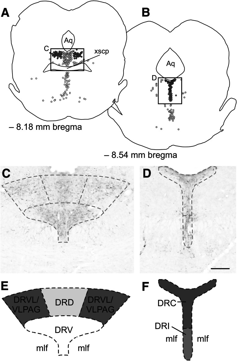
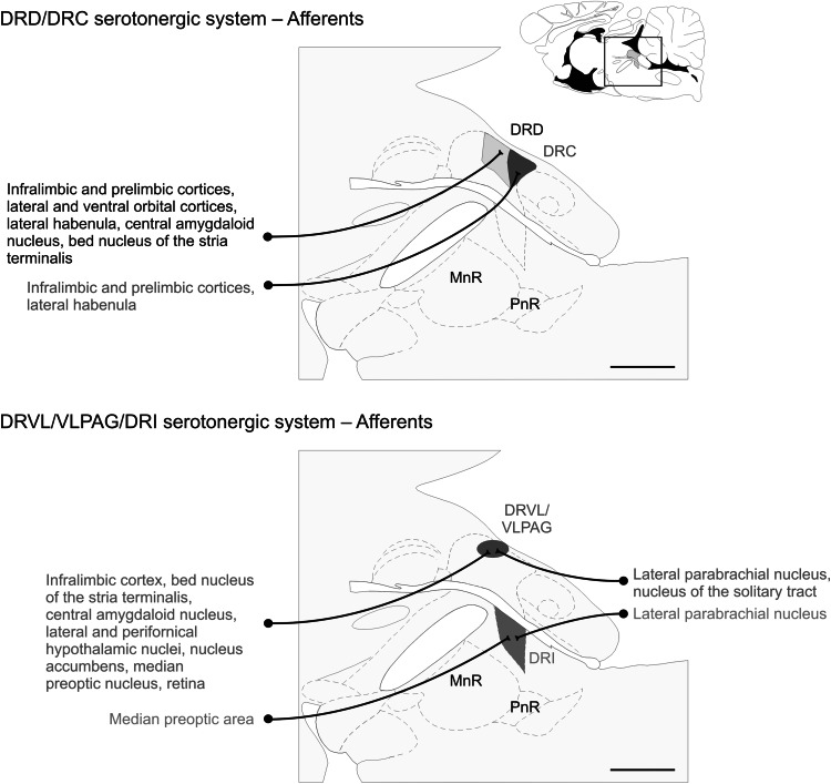
Previous studies have suggested that serotonergic neurons in the midbrain raphe complex have a functional topographic organization. Recent studies suggest that stimulation of a bed nucleus of the stria terminalis-dorsal raphe nucleus pathway by stress- and anxiety-related stimuli modulates a subpopulation of serotonergic neurons in the dorsal part of the dorsal raphe nucleus (DRD) and caudal part of the dorsal raphe nucleus (DRC) that participates in facilitation of anxiety-like responses. In contrast, recent studies suggest that activation of a spinoparabrachial pathway by peripheral thermal or immune stimuli excites subpopulations of serotonergic neurons in the ventrolateral part of the dorsal raphe nucleus/ventrolateral periaqueducal gray (DRVL/VLPAG) region and interfascicular part of the dorsal raphe nucleus (DRI). Studies support a role for serotonergic neurons in the DRVL/VLPAG in inhibition of panic-like responses, and serotonergic neurons in the DRI in antidepressant-like effects. Thus, data suggest that while some subpopulations of serotonergic neurons in the dorsal raphe nucleus play a role in facilitation of anxiety-like responses, others play a role in inhibition of anxiety- or panic-like responses, while others play a role in antidepressant-like effects. Understanding the anatomical and functional properties of these distinct serotonergic systems may lead to novel therapeutic strategies for the prevention and/or treatment of affective and anxiety disorders. In this review, we describe the anatomical and functional properties of subpopulations of serotonergic neurons in the dorsal raphe nucleus, with a focus on those implicated in symptoms of anxiety and affective disorders, the DRD/DRC, DRVL/VLPAG, and DRI.


Figures

Fig. 1
Fig. 1
The dorsal raphe nucleus can be divided into several subregions based on anatomical, functional, and hodological evidence. a Camera lucida drawing showing a coronal section of the rat midbrain/pons at −8.18 mm bregma. Each dot represents one serotonergic neuron in a 30 µm-thick section. An anxiety-related serotonergic system in the dorsal raphe nucleus, dorsal part (DRD) is represented by yellow dots, while an anti-panic serotonergic system in the dorsal raphe ventrolateral part/ventrolateral periaqueductal gray (DRVL/VLPAG) is represented by red dots. The black box in a is shown in the photomicrograph in c. c Photomicrograph showing tryptophan hydroxylase-immunoreactive neurons (orange/brown cytosolic staining) in the dorsal raphe nucleus at −8.18 mm bregma. Tryptophan hydroxylase (TPH) is the rate-limiting enzyme in the biosynthesis of serotonin and is commonly used as a marker of serotonergic neurons. Dashed lines show the boundaries for the subregions of the dorsal raphe nucleus which are depicted in e. e Schematic illustration showing the location of an anxiety-related serotonergic system in the DRD (yellow) and an anti-panic serotonergic system in the DRVL/VLPAG (red). b Camera lucida drawing showing a coronal section of the rat midbrain/pons at −8.54 mm bregma. Each dot represents one serotonergic neuron in a 30 µm-thick section. An anxiety-related serotonergic system in the dorsal raphe nucleus, caudal part (DRC) is represented by purple dots, while an antidepressant-like serotonergic system in the dorsal raphe nucleus, interfascicular part (DRI) is represented by green dots. The black box in b is shown in the photomicrograph in d. d Photomicrograph showing TPH-immunoreactive neurons in the dorsal raphe nucleus at −8.54 mm bregma. Dashed lines show the boundaries for the subregions of the dorsal raphe nucleus that are depicted in f. f Schematic illustration showing the location of an anxiety-related serotonergic system in the DRC (purple) and an antidepressant-like serotonergic system in the DRI (green)
Fig. 2
Fig. 2
The anxiety-related serotonergic system comprises the dorsal raphe nucleus, dorsal part (DRD), and dorsal raphe nucleus, caudal part (DRC) subregions of the dorsal raphe nucleus (DR), while the anti-panic/antidepressant serotonergic system comprises the dorsal raphe nucleus, ventrolateral part/ventrolateral periaqueductal gray (DRVL/VLPAG) and dorsal raphe nucleus, interfascicular part (DRI) subregions of the DR. These subregions receive topographically organized afferents from forebrain and brainstem regions. Schematic illustrations of midline sagittal sections of the rat brainstem show subdivisions of the DR adapted from a standard rat brain atlas (Paxinos and Watson 1998). The full sagittal section is shown in the top right corner for reference; the box shows the area of the brainstem illustrated in the main figure. The subdivisions of the DR and the forebrain and brainstem structures giving rise to selected prominent afferent projections to each subdivision are color-coordinated; DRC purple; DRD yellow/black text; DRI green; DRVL/VLPAG red. DRC dorsal raphe nucleus, caudal part; DRD dorsal raphe nucleus, dorsal part; DRI dorsal raphe nucleus, interfascicular part; DRVL/VLPAG dorsal raphe nucleus, ventrolateral part/ventrolateral periaqueductal gray; MnR median raphe nucleus; PnR pontine raphe nucleus. Scale bar, 1 mm
Fig. 3
Fig. 3
The anxiety-related serotonergic system comprises the dorsal raphe nucleus, dorsal part (DRD) and dorsal raphe nucleus, caudal part (DRC) subregions of the dorsal raphe nucleus (DR), while the anti-panic/antidepressant serotonergic system comprises the dorsal raphe nucleus, ventrolateral part/ventrolateral part of the periaqueductal gray (DRVL/VLPAG) and dorsal raphe nucleus, interfascicular part (DRI) subregions of the DR. These subregions send topographically organized projections to forebrain and brainstem regions via distinct serotonergic tracts. Schematic illustrations of midline sagittal sections of the rat brainstem show subdivisions of the DR adapted from a standard rat brain atlas (Paxinos and Watson 1998). The full sagittal section is shown in the top right corner for reference; the box shows the area of the brainstem illustrated in the main figure. There is an anatomic and functional topography among the different subregions of the DR. The lists on the left side of the figure indicate physiologic, pharmacologic, and behavioral stimuli known to activate serotonergic neurons in different subdivisions of the DR. The subdivisions of the DR, the ascending (Azmitia and Segal 1978) and descending serotonergic tracts, as well as the efferent projection regions are color-coordinated; DRC purple; DRD yellow/black text; DRI green; DRVL/VLPAG red. DRC dorsal raphe nucleus, caudal part; DRD dorsal raphe nucleus, dorsal part; DRI dorsal raphe nucleus, interfascicular part; DRVL/VLPAG dorsal raphe nucleus, ventrolateral part/ventrolateral periaqueductal gray; LPS lipopolysaccharide; MnR median raphe nucleus; PnR pontine raphe nucleus. Scale bar, 1 mm

References

    1. Abrams JK, Johnson PL, Hay-Schmidt A, Mikkelsen JD, Shekhar A, Lowry CA (2005) Serotonergic systems associated with arousal and vigilance behaviors following administration of anxiogenic drugs. Neuroscience 133:983–997 - PubMed
    1. Amaral DG, Cowan WM (1980) Subcortical afferents to the hippocampal formation in the monkey. J Comp Neurol 189:573–591 - PubMed
    1. Amat J, Tamblyn JP, Paul ED, Bland ST, Amat P, Foster AC, Watkins LR, Maier SF (2004) Microinjection of urocortin 2 into the dorsal raphe nucleus activates serotonergic neurons and increases extracellular serotonin in the basolateral amygdala. Neuroscience 129:509–519 - PubMed
    1. Amat J, Baratta MV, Paul E, Bland ST, Watkins LR, Maier SF (2005) Medial prefrontal cortex determines how stressor controllability affects behavior and dorsal raphe nucleus. Nat Neurosci 8:365–371 - PubMed
    1. Azmitia EC, Gannon PJ (1986) The primate serotonergic system: a review of human and animal studies and a report on Macaca fascicularis. In: Fahn S et al (eds) Myoclonus. Raven Press, New York, pp 407–468 - PubMed

Publication types