. 2012 Sep;15(3):409-20.
doi: 10.1007/s10456-012-9269-x. Epub 2012 Apr 7.

Inhibition of ARNT severely compromises endothelial cell viability and function in response to moderate hypoxia

Affiliations

Inhibition of ARNT severely compromises endothelial cell viability and function in response to moderate hypoxia

Yu Han et al. Angiogenesis. 2012 Sep.

Abstract

Hypoxia inducible factor (HIF) is a master heterodimeric transcriptional regulator of oxygen (O(2)) homeostasis critical to proper angiogenic responses. Due to the distinctive coexpression of HIF-1α and HIF-2α subunits in endothelial cells, our goal was to examine the genetic elimination of HIF transcriptional activity in response to physiological hypoxic conditions by using a genetic model in which the required HIF-β subunit (ARNT, Aryl hydrocarbon Receptor Nuclear Translocator) to HIF transcriptional responses was depleted. Endothelial cells (ECs) and aortic explants were isolated from Arnt ( loxP/loxP ) mice and infected with Adenovirus-Cre/GFP or control-GFP. We observed that moderate levels of 2.5 % O(2) promoted vessel sprouting, growth, and branching in control aortic ring assays while growth from Adenovirus-Cre infected explants was compromised. Primary Adenovirus-Cre infected EC cultures featured adverse migration and tube formation phenotypes. Primary pulmonary or cardiac ARNT-deleted ECs also failed to proliferate and survive in response to 8 or 2.5 % O(2) and hydrogen peroxide treatment. Our data demonstrates that ARNT promotes EC migration and vessel outgrowth and is indispensible for the proliferation and preservation of ECs in response to the physiological environmental cue of hypoxia. Thus, these results demonstrate that ARNT plays a critical intrinsic role in ECs and support an important collaboration between HIF-1 and HIF-2 transcriptional activity in these cells.


Figures

Fig. 1
Fig. 1
Loss of ARNT inhibits hypoxia enhanced vessel outgrowth and branching in aortic-ring explants. (A) Magnification of vessel outgrowth from rings stained for Rhodamine-CD31 (a, b). Note the vacuolated vessels in b (Arrowhead). Fluorescent micrographs of rings dissected from aortas of ArntloxP/loxP adult mice were infected with Ade -GFP (c) or -Cre/GFP (d). (B) Phase-contrast micrographs of representative rings infected with Ade -GFP or -Cre/GFP after culturing for 10 days under normoxic or hypoxic (2.5% O2) conditions. Arrows indicate vessel branches. Quantification of vascular outgrowth assessed by the number of vessels, length of vessels (C) and branches (D). Data shown are the mean ± SEM, n=10 samples. Statistical analysis was performed using an unpaired Student’s t-test.
Fig. 2
Fig. 2
Arnt-null endothelial cells fail to upregulate HIF target genes in response to hypoxia. Generation of Arnt-null endothelial cells (A and B). (A) PCR amplification of genomic DNA detecting ARNT deleted band. (B) Western blot analysis of ARNT protein from endothelial cells (EC4) cells derived from CD31+ICAM+ ECs isolated from lungs of ArntloxP/loxP mice which were then immortalized, infected with Adenovirus -GFP and -Cre/GFP, and FACS sorted. Proteins isolated from WT and Arnt-null (ArntΔ/Δ) embryonic stem cells were used as controls. (C) Real-time PCR analysis of HIF-target gene expression in control or Arnt-null EC4s and exposed to normoxic (21% O2) or hypoxic (8% or 2.5% O2) conditions for 16 hours. Relative transcript levels were all normalized to 18S RNA and compared to Ade-GFP normoxic EC4 cultures. Phosphoglycerate kinase 1 (PGK) is considered HIF-1α specific target; adenosine A2A receptor (Adora2a) and VEGF receptor -2 and -1 (Flk-1, and Flt-1) are considered HIF-2α specific targets; adrenomedullin (ADM) and vascular endothelial growth factor (VEGF), are regulated by both α subunits.
Fig. 3
Fig. 3
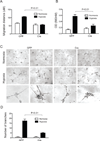
Loss of ARNT disrupts endothelial cell migration and tube formation. (A) Migration was detected in a 2-dimensional scratch wound assay of primary CD31+ICAM-2+ lung endothelial cells isolated from ArntloxP/loxP mice after 48 hours of infection with Adenovirus -GFP or -Cre/GFP. Migration was measured after 16 hours of normoxic and hypoxic (2.5% O2) treatment. Data are mean±SEM, n=3. (B) Cell migration capacity of immortalized EC4s examined in response to 15 ng/ml of VEGF using Boyden chamber cultures. The relative cell numbers remaining were quantified by measuring solubilized crystal violet uptake by cells remaining after removal of cells from upper side of chamber. Data are expressed as migrated cells and correspond to mean±SEM of 3 experiments performed in duplicate. (C) Representative pictures of control ArntloxP/loxP EC4 and Arnt-null EC4 cells plated on Matrigel for 24 hours. Panels: Upper, normoxic cultures; Middle, hypoxic cultures; Lower, higher magnification of the hypoxia panels. (D) Quantitative data of tube branch formation after 24 hours.
Figure 4
Figure 4
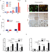
Arnt-null endothelial cells display proliferation and survival defects. ArntloxP/loxP primary lung and heart endothelial cells (ECs) were infected with Adenovirus -GFP or -Cre/GFP. (A) BrdU incorporation to assess lung EC proliferation under normoxic or hypoxic (2.5% O2) conditions. (B) Upper panels, Lysotracker Red (LTR) staining detecting cellular death in primary lung ECs. Arrows, LTR+/Cre+ cells. Middle/lower panels, TUNEL staining (brown) and quantification (C) of primary lung ECs cultures 24 hours following serum withdrawal in 2.5% or 21% O2 conditions. High magnification images are shown in the bottom right corner of each panel. (D,E) Quantification of Caspase-3 from primary lung (D) and heart (E) endothelial cells in response to 21, 8, or 2.5% O2for 24 hours or 300uM H2O2 for 2 hours.

Similar articles

Cited by

References

    1. Lonergan KM, Iliopoulos O, Ohh M, Kamura T, Conaway RC, Conaway JW, Kaelin WG., Jr Regulation of hypoxia-inducible mrnas by the von hippel-lindau tumor suppressor protein requires binding to complexes containing elongins b/c and cul2. Mol Cell Biol. 1998;18(2):732–741. - PMC - PubMed
    1. Cockman ME, Masson N, Mole DR, Jaakkola P, Chang GW, Clifford SC, Maher ER, Pugh CW, Ratcliffe PJ, Maxwell PH. Hypoxia inducible factor-alpha binding and ubiquitylation by the von hippel-lindau tumor suppressor protein. J Biol Chem. 2000;275(33):25733–25741. - PubMed
    1. Epstein JM, Gleadle AC, McNeill LA, Hewitson KS, O'Rourke J, Mole DR, Mukherji M, Metzen E, Wilson MI, Dhanda A, Tian YM, Masson N, Hamilton DL, Jaakkola P, Barstead R, Hodgkin J, Maxwell PH, Pugh CW, Schofield CJ, Ratcliffe PJ. C. Elegans egl-9 and mammalian homologs define a family of dioxygenases that regulate hif by prolyl hydroxylation. Cell. 2001;107(1):43–54. - PubMed
    1. Wang BH, Jiang GL, Rue EA, Semenza GL. Hypoxia-inducible factor 1 is a basic-helix-loop-helix-pas heterodimer regulated by cellular o2 tension. Proc Natl Acad Sci U S A. 1995;92(12):5510–5514. - PMC - PubMed
    1. Ramirez-Bergeron DL, Runge A, Adelman DM, Gohil M, Simon MC. Hif-dependent hematopoietic factors regulate the development of the embryonic vasculature. Dev Cell. 2006;11(1):81–92. - PMC - PubMed

Publication types

Substances

LinkOut - more resources