. 2012;7(4):e34202.
doi: 10.1371/journal.pone.0034202. Epub 2012 Apr 2.

Structural insights into TIR domain specificity of the bridging adaptor Mal in TLR4 signaling

Affiliations

Structural insights into TIR domain specificity of the bridging adaptor Mal in TLR4 signaling

Zhijie Lin et al. PLoS One. 2012.

Abstract

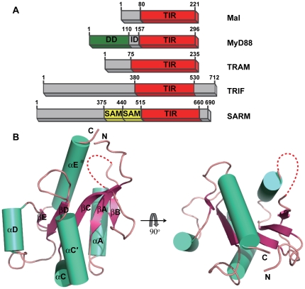
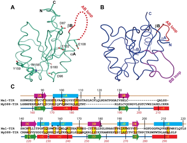
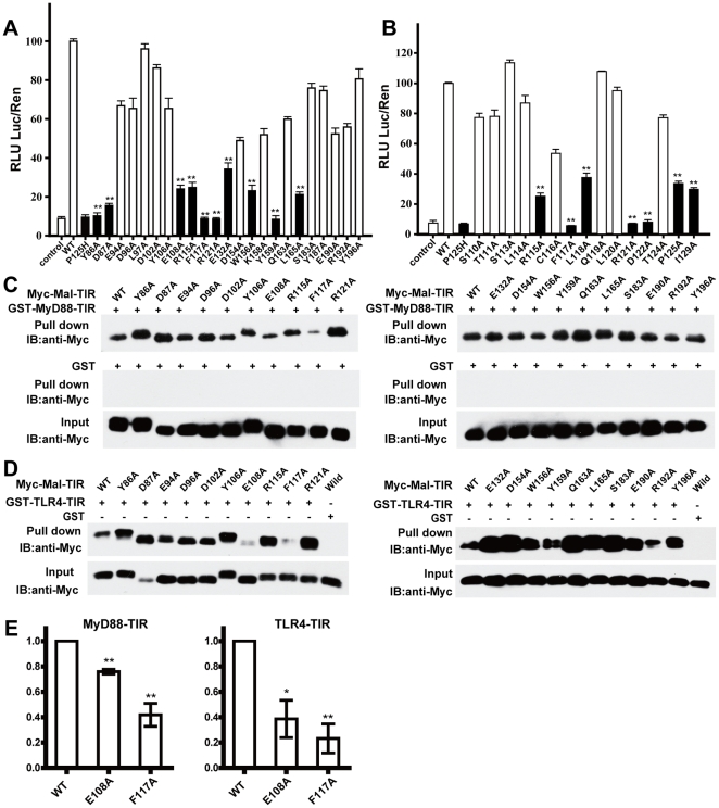
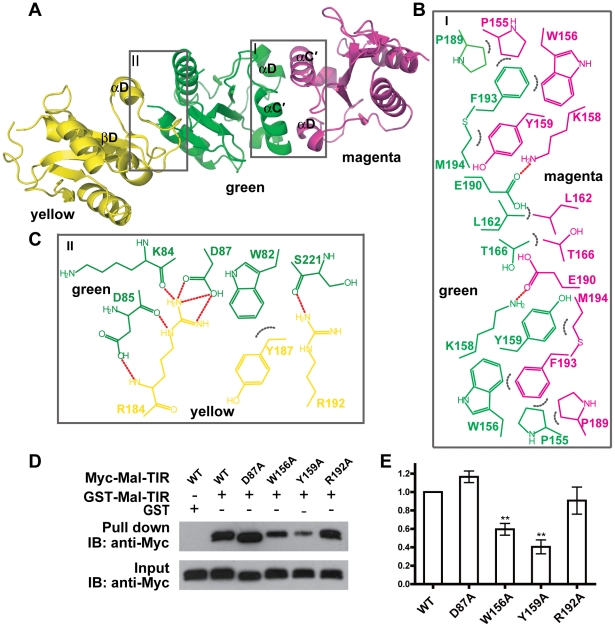
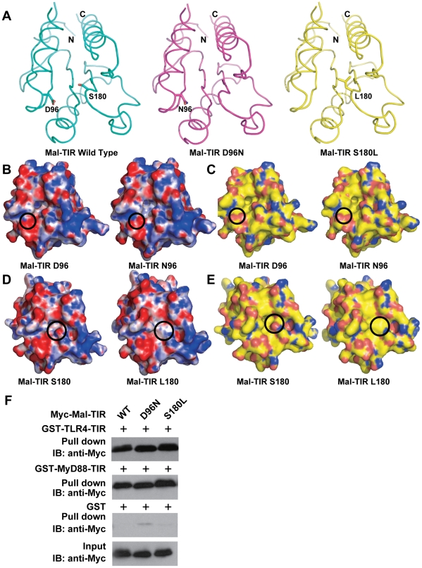
MyD88 adaptor-like protein (Mal) is a crucial adaptor that acts as a bridge to recruit the MyD88 molecule to activated TLR4 receptors in response to invading pathogens. The specific assembly of the Toll/interleukin-1 receptor (TIR) domains of TLR4, Mal and MyD88 is responsible for proper signal transduction in the TLR4 signaling pathway. However, the molecular mechanism for the specificity of these TIR domains remains unclear. Here, we present the crystal structure of the TIR domain of the human Mal molecule (Mal-TIR) at a resolution of 2.4 Å. Unexpectedly, Mal-TIR exhibits an extraordinarily long AB loop, but no αB helix or BB loop, distinguishing it from other TIR domains. More importantly, the Mal-TIR AB loop is capable of mediating direct binding to the TIR domains of TLR4 and MyD88 simultaneously. We also found that Mal-TIR can form a back-to-back dimer that may resemble the dimeric assembly of the entire Mal molecule. Our data demonstrate the bridge role of the Mal-TIR domain and provide important information about TIR domain specificity.


Conflict of interest statement

Competing Interests: The authors have declared that no competing interests exist.

Figures

Figure 1
Figure 1. Crystal structure of Mal-TIR.
(A) Schematic illustration of five TIR-domain-containing adaptors involved in TLR signaling. Mal: MyD88 adaptor-like protein; MyD88: myeloid differentiation factor 88; TRIF: TIR-domain-containing adaptor-inducing interferon-β; TRAM: TRIF-related adaptor molecule; SARM: sterile and HEAT/armadillo (ARM) motif protein; DD: death domain; ID: intermediate domain; SAM: sterile α-motif. (B) Cartoon representation of the structure of Mal-TIR. The β-strands are shown in pink, the α-helices in cyan and the connecting loops in salmon. The AB loop is colored red. The red dotted line represents the region that is not resolved in this structure.
Figure 2
Figure 2. Comparison of the structures of Mal-TIR and MyD88-TIR.
(A,B) Structural comparison of Mal (cyan) and MyD88 (blue). In Mal-TIR, the AB loop is colored red. In MyD88-TIR, the AB loop is shown in red and the BB loop in magenta. Residues D87, D96, E108, E132, W156, Y159 and S180 are shown. (C) Sequence alignment between human Mal-TIR and MyD88-TIR based on structural conservation. Note the differences in the region from αA to βC. Secondary structural elements of both proteins are shown as cylinders (α-helices) and arrows (β-strands). Highly conserved residues are highlighted in yellow, and residues that are similar across the group of sequences are boxed in red. Residues D87, D96, E108, F117, E132, W156, Y159 and S180 are labeled with asterisks.
Figure 3
Figure 3. Mutational analysis of the Mal-TIR domain.
(A) Selective mutants of solvent-exposed residues that are highly conserved across different species and used in the NF-κB reporter assay. (B) Alanine scanning of each residue of the AB loop (except alanine and glycine) for the NF-κB reporter assay. Black bars indicate decreased (less than 50%) NF-κB activity. RLU: relative luciferase unit; Luc: firefly luciferase activity; Ren: Renilla luciferase activity. GST pull-down assay for the MyD88-TIR and Mal-TIR variants (C) and for the TLR4-TIR and Mal-TIR variants (D). MyD88-TIR and TLR4-TIR were purified as GST fusion proteins. Mal-TIR was double-tagged with His6 and Myc and purified with Ni-NTA. The resulting complexes were analyzed by SDS-PAGE and Western blotting. (E) Bar graph of Mal-TIR wild type and mutants (E108A and F117A) binding to MyD88-TIR (left) or TLR4-TIR (right). The error bars indicate the standard error of the mean (n = 3 separate experiments). * indicates a P value<0.05, ** indicates a P value<0.001.
Figure 4
Figure 4. Dimeric packing of Mal-TIR.
(A) Ribbon representation of two possible interfaces related by crystallographic packing. Chemical drawing of the key residues involved in the interactions within the interface of the symmetric dimer (B) and the interface of the asymmetric dimer (C). Hydrogen bonds are shown as dotted red lines and hydrophobic interactions as dotted gray arcs. (D) GST pull-down assay for the homodimerization of Mal-TIR. Wild type Mal-TIR was purified as a GST fusion protein. Double-tagged (His6 and Myc) Mal-TIR wild type and mutants were purified with Ni-NTA. The resulting complexes were analyzed by SDS-PAGE and Western blotting. (E) Bar graph of Mal-TIR homodimerization. The error bars indicate the standard error of the mean (n = 3 separate experiments). ** indicates a P value<0.001.
Figure 5
Figure 5. Biochemical characteristics of wild-type and mutant Mal-TIR domains.
(A) Structural comparison between wild-type and mutant Mal-TIR domains. Crystal structures of the wild-type (cyan), D96N (magenta) and S180L (yellow) mutants of the Mal-TIR domain are shown. The residues Asp96 and Ser180 in the wild type as well as Asn96 in D96N and Leu180 in S180L are labeled. Nitrogen and oxygen atoms in the side chains are colored blue and red, respectively. (B,D) Electrostatic surfaces of wild type and the D96N mutant or the S180L mutant. Surfaces are colored by electrostatic potential ranging from red (−10 kbT/ec) to blue (+10 kbT/ec), where kb is the Boltzmann constant, T is temperature and ec is the electron charge. The electrostatic potentials were calculated by solving the Poisson-Boltzmann equation with APBS plugin of PyMol program (DeLano Scientific LLC). (C,E) The molecular surfaces of wild-type (left) and the D96N mutant (right) or the S180L mutant of the Mal-TIR domains. Carbon, nitrogen, oxygen and sulfur atoms are colored yellow, blue, red and orange, respectively. (F) GST pull-down assay for the interaction of wild-type Mal-TIR and mutants with TLR4-TIR and MyD88-TIR.

References

    1. Barton GM, Kagan JC. A cell biological view of Toll-like receptor function: regulation through compartmentalization. Nat Rev Immunol. 2009;9:535–542. - PMC - PubMed
    1. Akira S, Uematsu S, Takeuchi O. Pathogen recognition and innate immunity. Cell. 2006;124:783–801. - PubMed
    1. Jin MS, Kim SE, Heo JY, Lee ME, Kim HM, et al. Crystal structure of the TLR1-TLR2 heterodimer induced by binding of a tri-acylated lipopeptide. Cell. 2007;130:1071–1082. - PubMed
    1. Liu L, Botos I, Wang Y, Leonard JN, Shiloach J, et al. Structural basis of toll-like receptor 3 signaling with double-stranded RNA. Science. 2008;320:379–381. - PMC - PubMed
    1. Choe J, Kelker MS, Wilson IA. Crystal structure of human toll-like receptor 3 (TLR3) ectodomain. Science. 2005;309:581–585. - PubMed

Publication types

MeSH terms