. 2011 Dec;1(4):311-318.
doi: 10.1556/EuJMI.1.2011.4.7. Epub 2011 Dec 23.

THE ROLE OF SPECIFIC IgG AND COMPLEMENT IN COMBATING A PRIMARY MUCOSAL INFECTION OF THE GUT EPITHELIUM

Affiliations

THE ROLE OF SPECIFIC IgG AND COMPLEMENT IN COMBATING A PRIMARY MUCOSAL INFECTION OF THE GUT EPITHELIUM

C Belzer et al. Eur J Microbiol Immunol (Bp). 2011 Dec.

Abstract

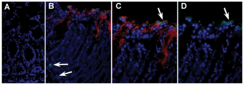
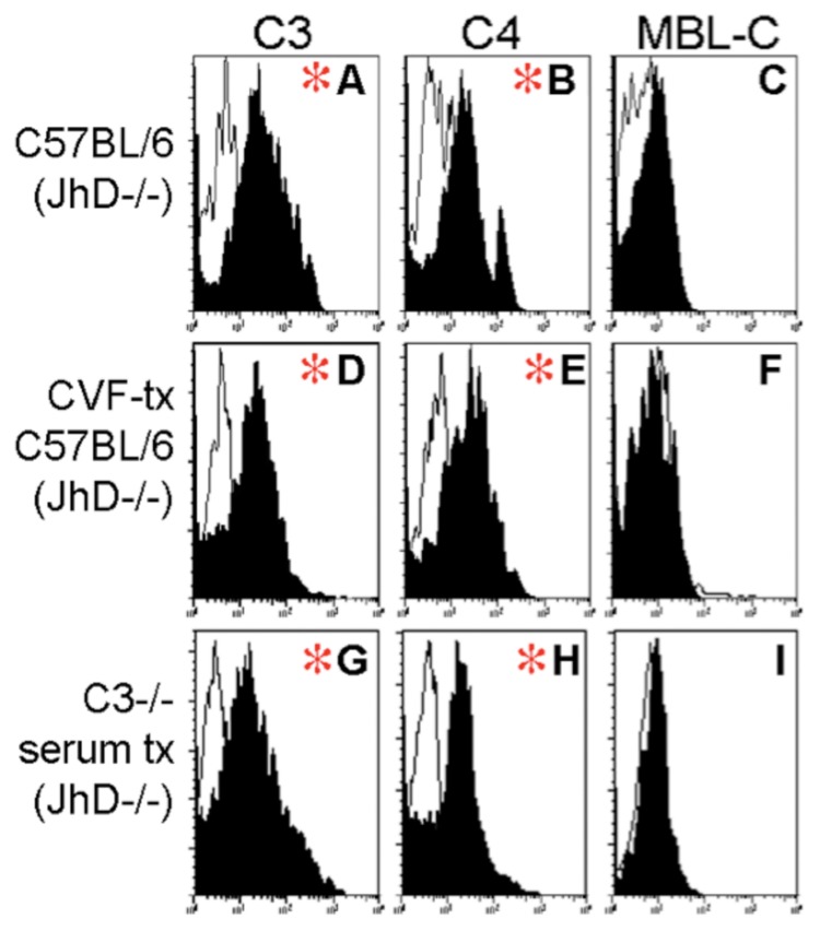
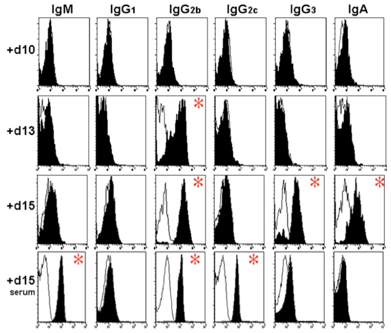
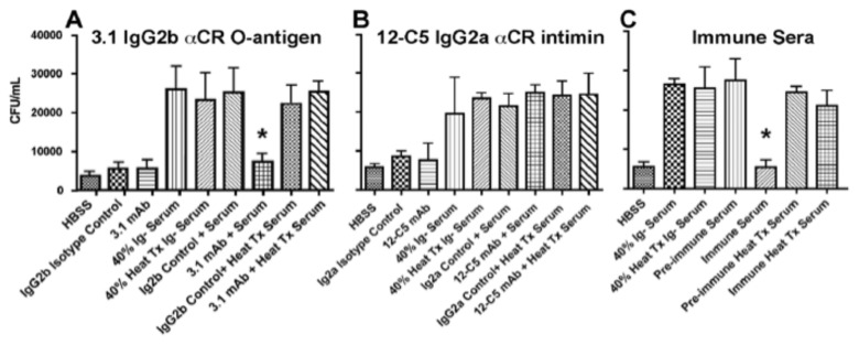
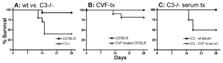
The role of complement and complement-fixing IgG isotypes at mucosal surfaces is ill defined. Previous data have demonstrated that survival of an infection with the attaching and effacing pathogen Citrobacter rodentium requires production of systemic and CD4+ T cell-dependent IgG. We have found that both complement and complement-fixing IgG isotypes are needed to survive a C. rodentium infection. Our results indicate that both IgG and complement C3b enter the gut lumen and bind epithelially adherent, and fecally shed C. rodentium. Furthermore, C3-deficient mice demonstrate a profound survival defect, though means to replenish C3 in systemic or mucosal sites restores the protective capacity of complement in the host. Our data provide evidence that both IgG and complement interact constructively on both sides of the epithelium to fight colonizing mucosal infections.


Figures

Fig. 1.
Fig. 1.
In vivo association of IgG and C3 with apically adherent C. rodentium: (A) Section of mouse colon (×200) stained with Hoescht dye donkey anti-rabbit-CY3 and rat-anti-mouse IgG-FITC. (B) Section from infected mouse (×200) showing host nuclei in blue, prominent C3 staining of apically adherent C. rodentium (red), faint IgG straining at the luminal surface (green), as well as IgG+ B cells in the lamina propria (arrows). (C) ×400 magnification of previous image showing distribution of C3+ C. rodentium and co-localization of IgG at the luminal surface. (D) Composite of previous image showing only IgG and Hoescht staining
Fig. 2.
Fig. 2.
Fixation of C3 on fecally shed C. rodentium: Representative histograms from flow cytometry of GFP+ events in fecal bacterial isolated from mice at 15 days of infection with C. rodentium. X-axis indicates fluorescence intensity in FL-2 (log10 scale); Y-axis indicates number of events. Panels A–C demonstrate staining in a representative wildtype mouse. (A) C3 and (B) C4, but not (C) MBL-C to shed GFP+CR (black plots). Staining in control antibody-deficient JhD–/– mice (white tracings) are shown. Red asterisk indicates populations where the mean fluorescence intensity between wildtype and JhD–/– mice was significant. Similar plots are shown from a representative CVF-treated mouse showing (D) C3, (E) C4, and (F) MBL-C. The bottom row of plots shows representative histograms from a C3–/– mouse reconstituted intravenously with wildtype serum containing C3. (G) C3, (H) C4, and (I) MBL-C
Fig. 3.
Fig. 3.
Ig Binding to fecal bacteria during infection: Representative histograms from flow cytometry of GFP+ events in fecal bacteria isolated from mice at 10, 13, and 15 days of infection with C. rodentium. X-axis indicates fluorescence intensity in FL-2 (log10 scale); Y-axis indicates number of events. Panels: Staining in control antibody-deficient JhD–/– mice mice (white tracings) are shown. From left to right, the different antibodies are shown (IgM, IgG1, IgG2b, IgG2c, IgG3, and IgA). The three top rows indicate levels in the fecal pellets 10, 13, and 15 days after infection. The bottom row shows antibody levels in serum 15 days after infection. Asterisks indicate a significant difference between wildtype and immune compromised mice
Fig. 4.
Fig. 4.
Anti-bacterial activities of mAbs with complement-sufficient serum: Assays for complement-dependent lysis after 2-h incubation with (A) 3.1 mAb against C. rodentium O-antigen; asterisk indicates significant growth inhibition of bacteria in the presence of serum and 5 µg of 3.1 mAb, (B) 12-C5 mAb against bacterial intimin, or (C) effects of immune and pre-immune serum on bacterial growth inhibition. Asterisk indicates growth inhibition in the presence immune serum, as compared with adjacent peaks measuring growth in pre-immune serum or in heat-treated sera
Fig. 5.
Fig. 5.
Survival studies in C3–/– and CVF-treated mice infected with C. rodentium: (A) Comparison of survival in wildtype (11 of 12 mice survived) and C3–/– mice (4 of 10 mice survived) infected with C. rodentium at 21 days of age. (B) Wildtype CVF (n = 10) or saline (n = 10) mice. (C) Infection in C3–/– mice treated with wildtype serum (n = 5) or CVF-treated serum (n = 4 mice)
Fig. 6.
Fig. 6.
Complement and antibody responses: Demonstrating antibody titer against C. rodentium Intimin, LPS and sonicate in wildtype (circles) and C3–/– (diamonds) 14 days post-inoculation with C. rodentium. From left to right: the different antibodies that were tested (IgM, IgG2b and IgG2c). From top to bottom: the three antigens (Intimin, LPS and sonicate). Asterisk indicates a significant difference between wildtype and C3–/–

References

    1. Wales AD, Woodward MJ, Pearson GR. Attaching-effacing bacteria in animals. J Comp Pathol. 2005 Jan;132(1):1–26. - PMC - PubMed
    1. Schauer DB, Falkow S. Attaching and effacing locus of a Citrobacter freundii biotype that causes transmissible murine colonic hyperplasia. Infect Immun. 1993 Jun;61(6):2486–2492. - PMC - PubMed
    1. Luperchio SA, Schauer DB. Molecular pathogenesis of Citrobacter rodentium and transmissible murine colonic hyperplasia. Microbes Infect. 2001 Apr;3(4):333–340. - PubMed
    1. Black RE, Cousens S, Johnson HL, Lawn JE, Rudan I, Bassani DG, Jha P, Campbell H, Walker CF, Cibulskis R, Eisele T, Liu L, Mathers C. Global, regional, and national causes of child mortality in 2008: a systematic analysis. Lancet. 2010 Jun 5;375(9730):1969–1987. - PubMed
    1. Campellone KG. Cytoskeleton-modulating effectors of enteropathogenic and enterohaemorrhagic Escherichia coli: Tir, EspFU and actin pedestal assembly. FEBS J. 2010 Jun;277(11):2390–2402. - PubMed

LinkOut - more resources