. 2012 May 22;6(5):4157-68.
doi: 10.1021/nn300652p. Epub 2012 Apr 13.

Enhanced delivery of chemotherapy to tumors using a multicomponent nanochain with radio-frequency-tunable drug release

Affiliations

Enhanced delivery of chemotherapy to tumors using a multicomponent nanochain with radio-frequency-tunable drug release

Pubudu M Peiris et al. ACS Nano. .

Abstract

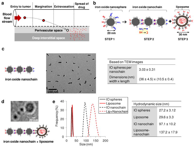
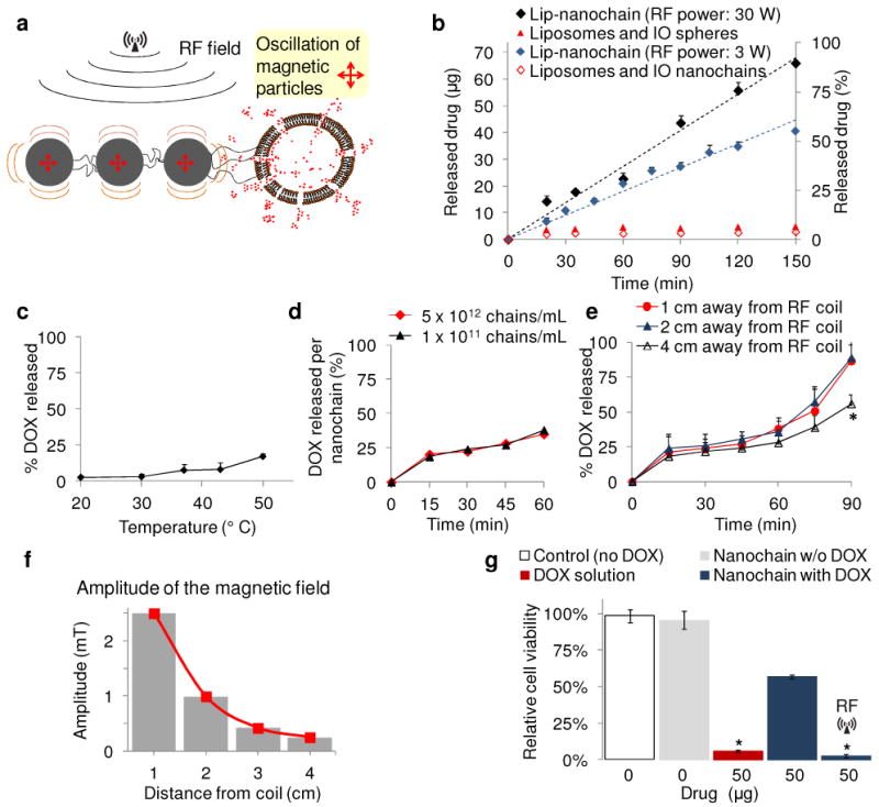
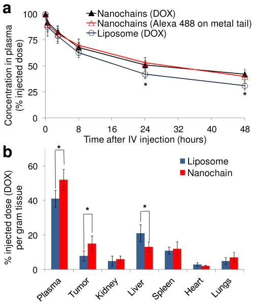
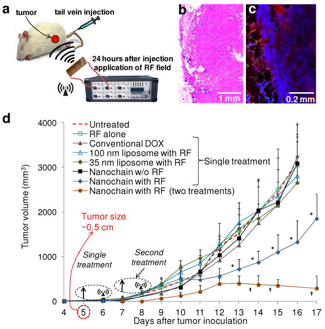
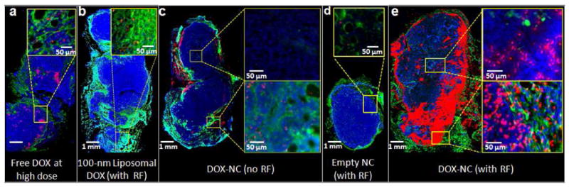
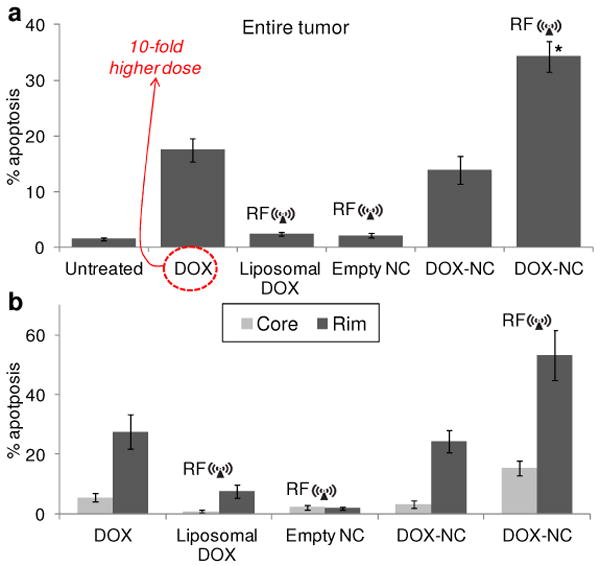
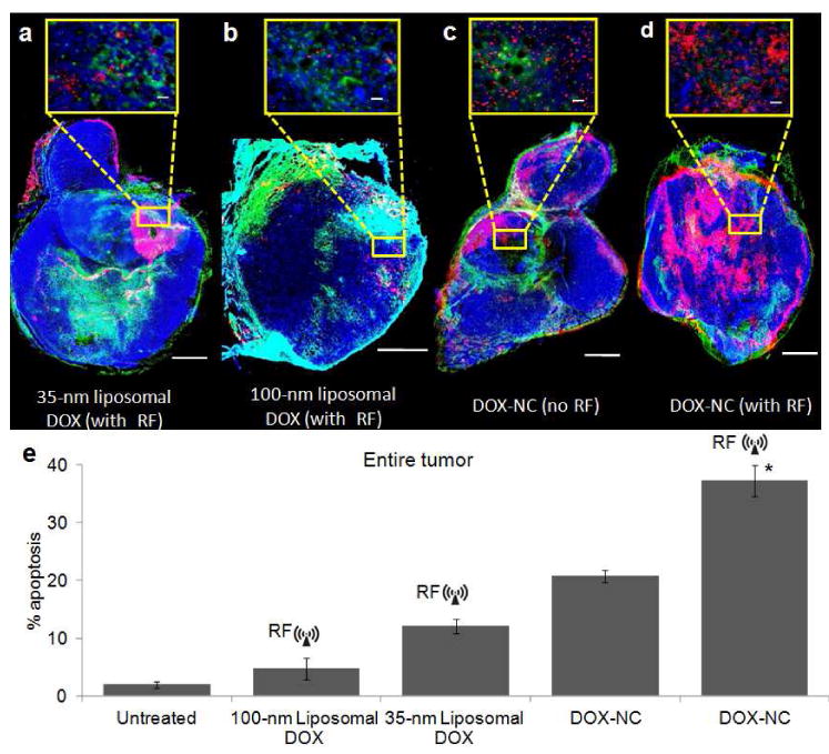
While nanoparticles maximize the amount of chemotherapeutic drug in tumors relative to normal tissues, nanoparticle-based drugs are not accessible to the majority of cancer cells because nanoparticles display patchy, near-perivascular accumulation in tumors. To overcome the limitations of current drugs in their molecular or nanoparticle form, we developed a nanoparticle based on multicomponent nanochains to deliver drug to the majority of cancer cells throughout a tumor while reducing off-target delivery. The nanoparticle is composed of three magnetic nanospheres and one doxorubicin-loaded liposome assembled in a 100 nm long chain. These nanoparticles display prolonged blood circulation and significant intratumoral deposition in tumor models in rodents. Furthermore, the magnetic particles of the chains serve as a mechanical transducer to transfer radio frequency energy to the drug-loaded liposome. The defects on the liposomal walls trigger the release of free drug capable of spreading throughout the entire tumor, which results in a widespread anticancer effect.


Figures

Fig. 1
Fig. 1
Characterization of the DOX-NC nanoparticle. (a) Illustration of the required steps for the successful delivery of nanoparticle-based drug to tumors. (b) Diagram of the DOX-NC nanoparticle and its constituent components including a nanochain composed of three iron oxide (IO) spheres and one liposome. (c) TEM image of magnetic nanochains composed of three IO spheres. The table summarizes the main characteristics of the magnetic nanochains obtained from visual analysis of TEM images (minimum count was 200 particles; data presented as mean ± s.d). (d) TEM image of a nanochain particle composed of three IO spheres and one DOX-loaded liposome. (e) Size distribution of the parent nanoparticles and DOX-loaded nanochains obtained by DLS measurements (data presented as mean ± s.d.)
Fig. 2
Fig. 2
In vitro evaluation of the RF-triggered release profile of DOX from DOX-NC particles. (a) Illustration of the defects on the liposome caused by ‘vibration’ of the IO spheres under an RF field. (b) Triggered release from DOX-NC particles using an RF field at 10 kHz and different energy outputs (the sample was located 1 cm away from the RF coil). The samples were exposed to the RF field for the entire duration of the experiment. Besides DOX-NC particles, the RF field (30 W) was applied to mixtures of liposomes with IO nanospheres or IO nanochains at a ratio of 1:3 (liposome: IO spheres). (c) Effect of temperature on the drug release from DOX-NC particles (incubation time was 60 min). (d) Drug release from DOX-NC particles at different particle concentration under an RF field at 10 kHz/30W (the sample was located 1 cm away from the RF coil). (e) Drug release from DOX-NC particles at different distance from the RF source (RF field: 10 kHz/30W). (f) Amplitude of the magnetic field at different distances from the RF source (RF field: 10 kHz/30W). (g) Cytotoxicity of DOX-NC (with or without RF) on 13762 MAT B III cells. Control treatments included black nanochains, free DOX, and liposomal DOX. The two data points marked with asterisks are statistically different compared to the other conditions (P<0.01).
Fig. 3
Fig. 3
Blood circulation and organ distribution of the DOX-NC particles in rats. (a) Plasma clearance of DOX-loaded liposomes (100 nm in diameter) and DOX-NC in rats at a dose of 0.5 mg/kg DOX (n=5). Besides DOX, fluorescence spectroscopy was used to measure Alexa 488 on the iron oxide particles (*P<0.05). (b) Organ and tumor distribution 24 h after administration of the DOX-loaded liposomes and DOX-NC at a dose of 0.5 mg DOX/kg in the rat 13762 MAT B III tumor model (n=6; *P<0.05).
Fig. 4
Fig. 4
In vivo treatment of breast tumor-bearing rats using DOX-NC particles. (a) Schematic of the therapeutic protocol. (b) Histological evaluation of the distribution of systemically administered DOX-NC particles (blue: Prussian blue stain) in a tumor. (c) Application of an RF field released DOX molecules (red) that localized in the nuclei of cancer cells (blue: DAPI). (d) Measurement of the tumor growth of 13763 MAT B III tumors in rats after systemic administration of DOX-NC at a dose of 0.5 mg/kg DOX (arrow; day 5) followed by application of the RF field (day 6). Control treatments included saline (untreated), RF alone, free DOX, 100-nm liposomal DOX (with RF), 35-nm liposomal DOX (with RF) and DOX-NC (without RF). Another group of animals received a second injection of DOX-NC (arrow; days 7) followed by RF application (day 8). Data points marked with asterisks are statistically significant relative to all the other single-treated groups. Data points marked with crosses are statistically significant relative to all groups (n=6; * and † P<0.05).
Fig. 5
Fig. 5
Histological evaluation of the apoptotic effect of DOX-NC in the rat MAT B III model. (a) Fluorescence image of a histological section of a tumor 48 h after IV injection of free DOX at 5 mg/kg. The specific endothelial antigen CD31 was stained (green). Nuclei (blue) were stained with DAPI. Apoptotic cell nuclei were stained with TUNEL (red). (b) No significant apoptosis was observed in a tumor 48 h after systemic administration of 100-nm liposomal DOX at 0.5 mg/kg (RF was applied 24 h after injection). (c) Few apoptotic cells were found in a tumor 48 h after systemic administration of DOX-NC at 0.5 mg/kg. (d) Negligible apoptosis was found in a tumor 48 h after systemic administration of an empty nanochain (RF was applied 24 h after injection). (e) A significant number of apoptotic cells was found in a tumor 48 h after systemic administration of liposomal DOX at 0.5 mg/kg (RF was applied 24 h after injection).
Fig. 6
Fig. 6
Quantitative histological evaluation of apoptosis in the rat MAT B III model. (a) A quantitative analysis of the fluorescence images was performed by comparing the total number of cancer and apoptotic cells of an entire tumor as measured in at least 20 histological sections per tumor (about 10,000 cells per section). The apoptotic effect on tumors treated with DOX-NC followed by RF was compared to the other DOX-based treatments (n=3 rats per group; * P<0.01). (b) Regional apoptosis in the tumor was measured based on the degree of vascularization. Using the endothelial cells staining (CD31), the well-vascularized rim of the tumor was distinguished from its core.
Fig. 7
Fig. 7
Histological evaluation of apoptosis in the mouse 4T1 model. (a) Fluorescence image of a histological section of a tumor 48 h after IV injection of 35-nm liposomal DOX at 0.5 mg/kg (CD31: green, DAPI: blue, TUNEL: red). RF was applied 24 h after injection. The scale bar is 1 mm (scale bar of the inset is 50 μm) (b) No significant apoptosis was observed in a tumor 48 h after systemic administration of 100-nm liposomal DOX at 0.5 mg/kg (RF was applied 24 h after injection). (c) More apoptotic cells were found in a tumor 48 h after systemic administration of DOX-NC at 0.5 mg/kg. (d) A significant number of apoptotic cells was found in a tumor 48 h after systemic administration of DOX-NC at 0.5 mg/kg followed by RF application 24 h after injection. (e) A quantitative analysis of apoptosis was performed by comparing the total number of cancer and apoptotic cells of an entire tumor (minimum 20 histological sections per tumor; n=3 mice per group; * P<0.01).

References

    1. Von Hoff DD, Layard MW, Basa P, Davis HL, Jr, Von Hoff AL, Rozencweig M, Muggia FM. Risk Factors for Doxorubicin-Induced Congestive Heart Failure. Ann Intern Med. 1979;91:710–717. - PubMed
    1. Maeda H, Wu J, Sawa T, Matsumura Y, Hori K. Tumor Vascular Permeability and the EPR Effect in Macromolecular Therapeutics: A Review. J Control Release. 2000;65:271–284. - PubMed
    1. Lasic DD. Doxorubicin in Sterically Stabilized Liposomes. Nature. 1996;380:561–562. - PubMed
    1. Dreher MR, Liu W, Michelich CR, Dewhirst MW, Yuan F, Chilkoti A. Tumor Vascular Permeability, Accumulation, and Penetration of Macromolecular Drug Carriers. J Natl Cancer Inst. 2006;98:335–344. - PubMed
    1. Yuan F. Transvascular Drug Delivery in Solid Tumors. Semin Radiat Oncol. 1998;8:164–175. - PubMed

Publication types

Substances