Review
. 2012 Oct;22(5):747-53.
doi: 10.1016/j.conb.2012.03.006. Epub 2012 Apr 7.

OSVZ progenitors in the human cortex: an updated perspective on neurodevelopmental disease

Affiliations
Review

OSVZ progenitors in the human cortex: an updated perspective on neurodevelopmental disease

Bridget E LaMonica et al. Curr Opin Neurobiol. 2012 Oct.

Abstract

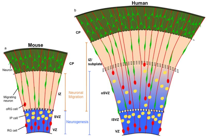
Recent discoveries concerning the architecture and cellular dynamics of the developing human brain are revealing new differences between mouse and human cortical development. In mice, neurons are produced by ventricular radial glial (RG) cells and subventricular zone intermediate progenitor (IP) cells. In the human cortex, both ventricular RG and highly motile outer RG cells generate IP cells, which undergo multiple rounds of transit amplification in the outer subventricular zone before producing neurons. This creates a more complex environment for neurogenesis and neuronal migration, adding new arenas in which neurodevelopmental disease gene mutation could disrupt corticogenesis. A more complete understanding of disease mechanisms will involve use of emerging model systems with developmental programs more similar to that of the human neocortex.


Figures

Figure 1
Figure 1. Increased spatial and cellular complexity of the developing human cortex
(A) Current model of mouse neocortical development. Primary zones of neurogenesis (blue) are the VZ and SVZ, where RG cells and IP cells reside, respectively. Neurons born in the VZ or SVZ migrate along the RG radial fiber scaffold found in the intermediate zone (IZ), the primary zone of neuronal migration (orange) in the mouse, to reach the CP. oRG cells are infrequent and not located in a distinct progenitor zone. (B) Expanded model of human neocortical development. RG cells, IP cells, and oRG cells are found in neurogenic zones (blue), which are the VZ, the inner SVZ (iSVZ), and the outer SVZ (oSVZ). Neurons migrate through the oSVZ and IZ/subplate (zones of neuronal migration, orange) to populate the CP. Neurons must navigate a larger distance than in the mouse, and a radial fiber scaffold of increased complexity, to reach the CP.
Figure 2
Figure 2. Molecular mechanisms of centrosome and microtubule-related cell behaviors in the developing neocortex
(A) An RG cell is depicted undergoing apical-to-basal INM during the G1 phase of the cell cycle. The basal direction is up, apical is down, and the centrosome (red) is located at the ventricular surface at the base of the primary cilium (not shown). Kinesin and actomyosin motors control nuclear movement, which is ab-centrosomal and toward the microtubule plus ends. (B) RG cell undergoing basal-to-apical INM during the G2 phase of the cell cycle. Nuclear movement is ad-centrosomal and towards microtubule minus ends. The process is controlled by dynein and associated proteins such as Lis1, and possibly by actomyosin motors. The centrosome has already replicated at this time. (C) RG cell division at the ventricular surface. The centrosomes move to opposite poles of the cell and function throughout mitosis in microtubule nucleation, spindle and microtubule attachment to kinetochores, cytokinesis, and mitotic exit. The centrosome also plays a role in asymmetric inheritance of cell fate components in daughter cells. (D) Radial neuronal migration along an RG fiber. The centrosome moves into the leading process prior to nucleokinesis, and a microtubule lattice originating from the centrosome forms a cage around the nucleus. Dynein and associated proteins function to pull the nucleus into the leading process. (E) oRG cell mitotic somal translocation (MST) is depicted with possible molecular mechanisms for controlling this cell behavior. Analogous to neuronal migration, movement of the centrosome precedes the nucleus into the basal process; therefore MST likely involves nuclear movement toward microtubule minus ends. MST may utilize minus-end directed motors such as dynein, and/or actomyosin motors.

References

    1. Hansen DV, Lui JH, Parker PR, Kriegstein AR. Neurogenic radial glia in the outer subventricular zone of human neocortex. Nature. 2010;464(7288):554–61. Using histological labeling techniques and timelapse imaging, this paper describes the progenitor cell composition of the developing human neocortex. The authors show that a novel class of radial glia found in the OSVZ, termed oRG cells, are neural stem cells, and may be responsible for increasing neuronal number and cortical gyration in evolution. - PubMed
    1. Fietz SA, Kelava I, Vogt J, Wilsch-Brauninger M, Stenzel D, Fish JL, Corbeil D, Riehn A, Distler W, Nitsch R, Huttner WB. OSVZ progenitors of human and ferret neocortex are epithelial-like and expand by integrin signaling. Nat Neurosci. 2010;13(6):690–9. This paper describes the presence of oRG cells in the human, and extends analysis of oRG cells analysis to another gyrencephalic mammal, the ferret. This is the first evidence that oRG cells are not specific to humans or primates, and may represent a more general mechanism for cortical expansion in many species. - PubMed
    1. Martinez-Cerdeno V, Cunningham CL, Camacho J, Antczak JL, Prakash AN, Cziep ME, Walker AI, Noctor SC. Comparative analysis of the subventricular zone in rat, ferret and macaque: evidence for an outer subventricular zone in rodents. PLoS One. 2012;7(1):e30178. - PMC - PubMed
    1. Kelava I, Reillo I, Murayama AY, Kalinka AT, Stenzel D, Tomancak P, Matsuzaki F, Lebrand C, Sasaki E, Schwamborn JC, Okano H, Huttner WB, Borrell V. Abundant Occurrence of Basal Radial Glia in the Subventricular Zone of Embryonic Neocortex of a Lissencephalic Primate, the Common Marmoset Callithrix jacchus. Cereb Cortex. 2012;22(2):469–81. - PMC - PubMed
    1. Garcia-Moreno F, Vasistha NA, Trevia N, Bourne JA, Molnar Z. Compartmentalization of cerebral cortical germinal zones in a lissencephalic primate and gyrencephalic rodent. Cereb Cortex. 2012;22(2):482–92. - PubMed

Publication types