

Epigenetic rejuvenation
- PMID: 22487104
- PMCID: PMC3444684
- DOI: 10.1111/j.1365-2443.2012.01595.x
Epigenetic rejuvenation
Abstract
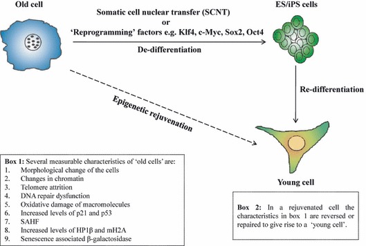
Induced pluripotent stem (iPS) cells have provided a rational means of obtaining histo-compatible tissues for 'patient-specific' regenerative therapies (Hanna et al. 2010; Yamanaka & Blau 2010). Despite the obvious potential of iPS cell-based therapies, there are certain problems that must be overcome before these therapies can become safe and routine (Ohi et al. 2011; Pera 2011). As an alternative, we have recently explored the possibility of using 'epigenetic rejuvenation', where the specialized functions of an old cell are rejuvenated in the absence of any change in its differentiated state (Singh & Zacouto 2010). The mechanism(s) that underpin 'epigenetic rejuvenation' are unknown and here we discuss model systems, using key epigenetic modifiers, which might shed light on the processes involved. Epigenetic rejuvenation has advantages over iPS cell techniques that are currently being pursued. First, the genetic and epigenetic abnormalities that arise through the cycle of dedifferentiation of somatic cells to iPS cells followed by redifferentiation of iPS cells into the desired cell type are avoided (Gore et al. 2011; Hussein et al. 2011; Pera 2011): epigenetic rejuvenation does not require passage through the de-/redifferentiation cycle. Second, because the aim of epigenetic rejuvenation is to ensure that the differentiated cell type retains its specialized function it makes redundant the question of transcriptional memory that is inimical to iPS cell-based therapies (Ohi et al. 2011). Third, to produce unrelated cell types using the iPS technology takes a long time, around three weeks, whereas epigenetic rejuvenation of old cells will take only a matter of days. Epigenetic rejuvenation provides the most safe, rapid and cheap route to successful regenerative medicine.
© 2012 The Authors. Journal compilation © 2012 by the Molecular Biology Society of Japan/Blackwell Publishing Ltd.
Figures
References
-
- Adams PD. Healing and hurting: molecular mechanisms, functions, and pathologies of cellular senescence. Mol. Cell. 2009;36:2–14. - PubMed
-
- Bahar R, Hartmann CH, Rodriguez KA, Denny AD, Busuttil RA, Dolle ME, Calder RB, Chisholm GB, Pollock BH, Klein CA, Vijg J. Increased cell-to-cell variation in gene expression in ageing mouse heart. Nature. 2006;441:1011–1014. - PubMed
-
- Billur M, Bartunik HD, Singh PB. The essential function of HP1 beta: a case of the tail wagging the dog? Trends Biochem. Sci. 2010;35:115–123. - PubMed
-
- Campisi J, d’Adda di Fagagna F. Cellular senescence: when bad things happen to good cells. Nat. Rev. Mol. Cell Biol. 2007;8:729–740. - PubMed
Publication types
MeSH terms
Substances
LinkOut - more resources
Full Text Sources
Other Literature Sources
Research Materials
