Review
. 2012 Aug;51(2):241-8.
doi: 10.1016/j.bone.2012.03.023. Epub 2012 Mar 28.

Effects of aging on articular cartilage homeostasis

Affiliations
Review

Effects of aging on articular cartilage homeostasis

Martin Lotz et al. Bone. 2012 Aug.

Abstract

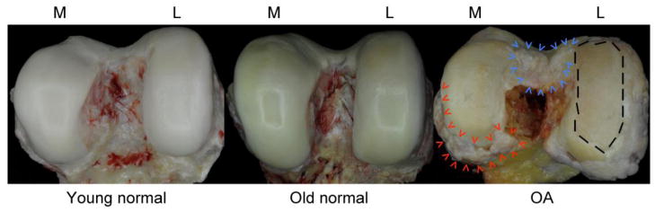
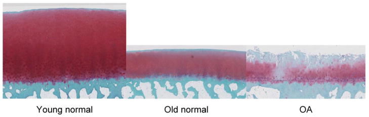
This review is focused on aging-related changes in cells and extracellular matrix of the articular cartilage. Major extracellular matrix changes are a reduced thickness of cartilage, proteolysis, advanced glycation and calcification. The cellular changes include reduced cell density, cellular senescence with abnormal secretory profiles, and impaired cellular defense mechanisms and anabolic responses. The extracellular and cellular changes compound each other, leading to biomechanical dysfunction and tissue destruction. The consequences of aging-related changes for joint homeostasis and risk for osteoarthritis are discussed. This article is part of a Special Issue entitled "Osteoarthritis".


Figures

Fig. 1
Fig. 1. Macroscopic images of human femoral condyles
Images represent normal young (left, age 40), normal aging (center, age 76) OA (right, age 88) tissue. The old normal condyle shows intact cartilage with yellow discoloration, which is in part due to the formation of advanced glycation end products. The OA sample features large areas with complete loss of articular cartilage (dashed line on the left femoral condyle), osteophytes at the joint margins (red arrowheads) and the intercondylar notch (blue arrowheads). M=medial; L=lateral
Fig. 2
Fig. 2. Safranin O stained sections of human femoral condyles
Images show young normal (left, age 40), old normal (center, age 76) and OA donors (right, age 88). The old normal sample has reduced tissue height, reduced safranin O staining in the superficial zone but no structural defects at the surface. The OA sample shows loss the superficial and part of the mid zone, fibrillations extending into the deep zone, areas with low cell density and cell clusters and duplication of the tidemark. All images are shown at 2.5x magnification.

References

    1. Zhang Y, Jordan JM. Epidemiology of osteoarthritis. Clin Geriatr Med. 2010;26:355–69. - PMC - PubMed
    1. Murphy L, Schwartz TA, Helmick CG, Renner JB, Tudor G, Koch G, Dragomir A, Kalsbeek WD, Luta G, Jordan JM. Lifetime risk of symptomatic knee osteoarthritis. Arthritis Rheum. 2008;59:1207–13. - PMC - PubMed
    1. Oliveria SA, Felson DT, Reed JI, Cirillo PA, Walker AM. Incidence of symptomatic hand, hip, and knee osteoarthritis among patients in a health maintenance organization. Arthritis Rheum. 1995;38:1134–41. - PubMed
    1. Hashimoto S, Ochs RL, Komiya S, Lotz M. Linkage of chondrocyte apoptosis and cartilage degradation in human osteoarthritis. Arthritis and Rheumatism. 1998;41:1632–8. - PubMed
    1. Temple-Wong MM, Bae WC, Chen MQ, Bugbee WD, Amiel D, Coutts RD, Lotz M, Sah RL. Biomechanical, structural, and biochemical indices of degenerative and osteoarthritic deterioration of adult human articular cartilage of the femoral condyle. Osteoarthritis Cartilage. 2009;17:1469–76. - PMC - PubMed

Publication types