

Isoflurane delays the development of early brain injury after subarachnoid hemorrhage through sphingosine-related pathway activation in mice
- PMID: 22488000
- PMCID: PMC3358576
- DOI: 10.1097/CCM.0b013e3182474bc1
Isoflurane delays the development of early brain injury after subarachnoid hemorrhage through sphingosine-related pathway activation in mice
Abstract
Objective: Isoflurane, a volatile anesthetic agent, has been recognized for its potential neuroprotective properties and has antiapoptotic effects. We examined whether isoflurane posttreatment is protective against early brain injury after subarachnoid hemorrhage and determined whether this effect needs sphingosine-related pathway activation.
Design: Controlled in vivo laboratory study.
Setting: Animal research laboratory.
Subjects: One hundred seventy-nine 8-wk-old male CD-1 mice weighing 30-38 g.
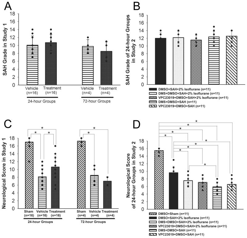
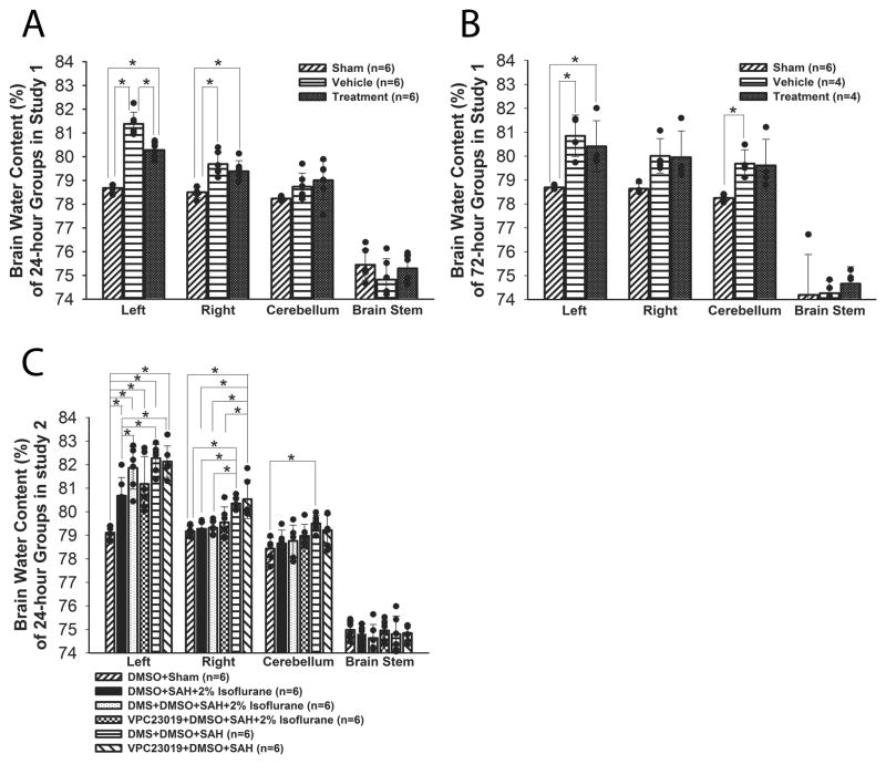
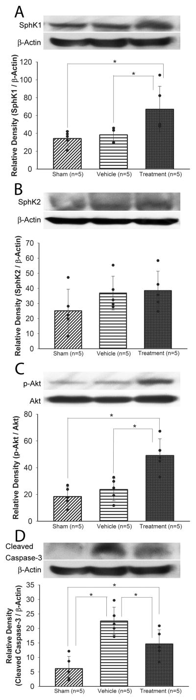
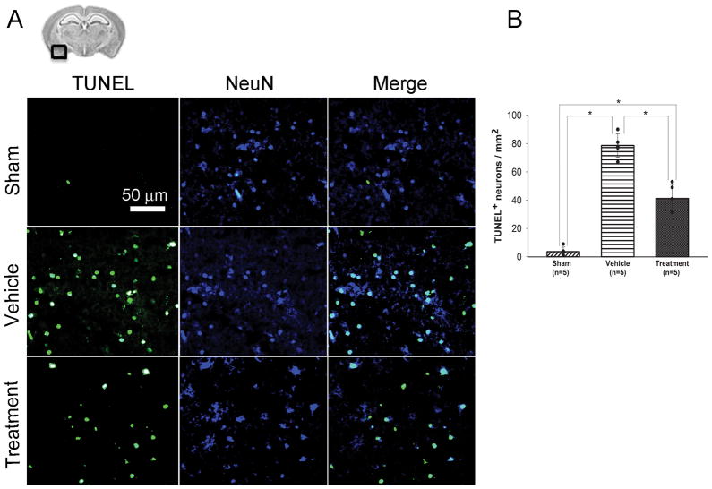
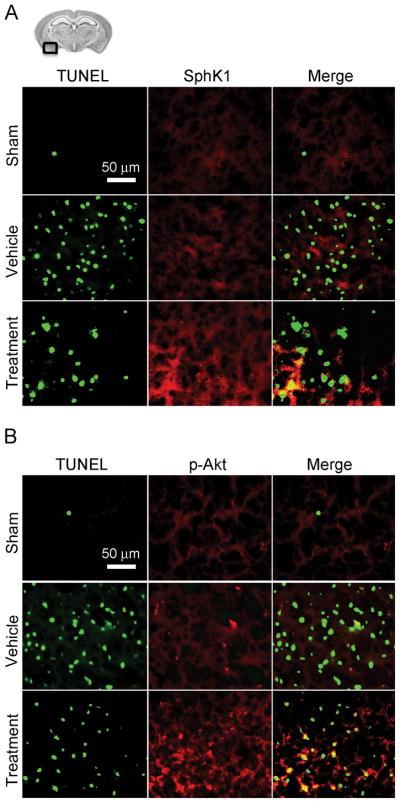
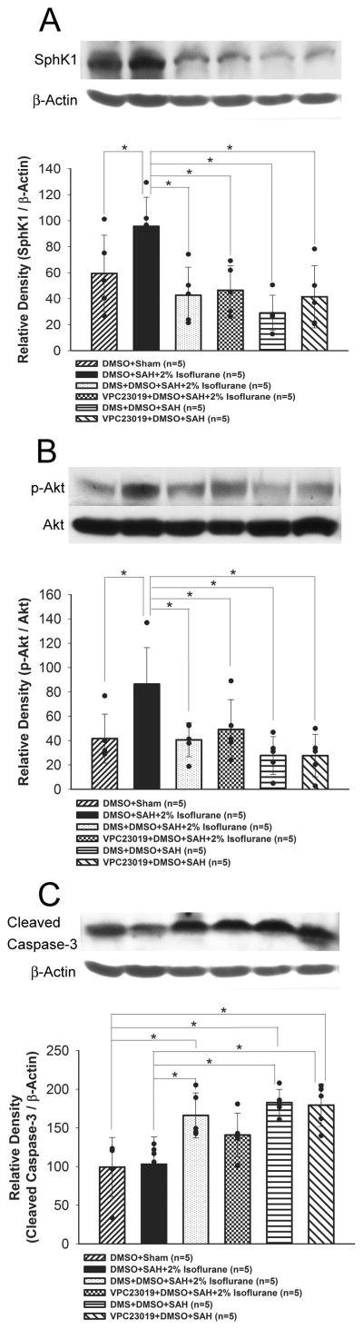
Interventions: Subarachnoid hemorrhage was induced in mice by endovascular perforation. Animals were randomly assigned to sham-operated, subarachnoid hemorrhage-vehicle, and subarachnoid hemorrhage+2% isoflurane. Neurobehavioral function and brain edema were evaluated at 24 and 72 hrs. The expression of sphingosine kinase, phosphorylated Akt, and cleaved caspase-3 was determined by Western blotting and immunofluorescence. Neuronal cell death was examined by terminal deoxynucleotidyl transferase-mediated uridine 5'-triphosphate-biotin nick end-labeling staining. Effects of a sphingosine kinase inhibitor N, N-dimethylsphingosine or a sphingosine 1 phosphate receptor inhibitor VPC23019 on isoflurane's protective action against postsubarachnoid hemorrhage early brain injury were also examined.
Measurements and main results: Isoflurane significantly improved neurobehavioral function and brain edema at 24 hrs but not 72 hrs after subarachnoid hemorrhage. At 24 hrs, isoflurane attenuated neuronal cell death in the cortex, associated with an increase in sphingosine kinase 1 and phosphorylated Akt, and a decrease in cleaved caspase-3. The beneficial effects of isoflurane were abolished by N, N-dimethylsphingosine and VPC23019.
Conclusions: Isoflurane posttreatment delays the development of postsubarachnoid hemorrhage early brain injury through antiapoptotic mechanisms including sphingosine-related pathway activation, implying its use for anesthesia during acute aneurysm surgery or intervention.
Conflict of interest statement
The authors have not disclosed any potential conflict of interest.
Figures
Comment in
-
Volatile meets versatile: does isoflurane strike a new path to neuroprotection?Crit Care Med. 2012 Jun;40(6):1992-3. doi: 10.1097/CCM.0b013e31824c9034. Crit Care Med. 2012. PMID: 22610220 No abstract available.
References
-
- Van Gijn J, Rinkel GJ. Subarachnoid haemorrhage: diagnosis, causes and management. Brain. 2001;124:249–278. - PubMed
-
- Maceyka M, Payne SG, Milstein S, et al. Sphingosine kinase, sphingosine-1-phosphate, and apoptosis. Biochim Biophys Acta. 2002;1585:193–201. - PubMed
-
- Dev KK, Mullerhausen F, Mattes H, et al. Brain sphingosine-1-phosphate receptors: implication for FTY720 in the treatment of multiple sclerosis. Pharmacol Ther. 2008;117:77–93. - PubMed
-
- Antkowiak B. How do general anaesthetics work? Naturwissenschaften. 2001;88:201–213. - PubMed
Publication types
MeSH terms
Substances
Grants and funding
LinkOut - more resources
Full Text Sources
Other Literature Sources
Research Materials
