Review
. 2012 Jun;69(6):337-70.
doi: 10.1002/cm.21031. Epub 2012 May 4.

Tropomodulins: pointed-end capping proteins that regulate actin filament architecture in diverse cell types

Affiliations
Review

Tropomodulins: pointed-end capping proteins that regulate actin filament architecture in diverse cell types

Sawako Yamashiro et al. Cytoskeleton (Hoboken). 2012 Jun.

Abstract

Tropomodulins are a family of four proteins (Tmods 1-4) that cap the pointed ends of actin filaments in actin cytoskeletal structures in a developmentally regulated and tissue-specific manner. Unique among capping proteins, Tmods also bind tropomyosins (TMs), which greatly enhance the actin filament pointed-end capping activity of Tmods. Tmods are defined by a TM-regulated/Pointed-End Actin Capping (TM-Cap) domain in their unstructured N-terminal portion, followed by a compact, folded Leucine-Rich Repeat/Pointed-End Actin Capping (LRR-Cap) domain. By inhibiting actin monomer association and dissociation from pointed ends, Tmods regulate actin dynamics and turnover, stabilizing actin filament lengths and cytoskeletal architecture. In this review, we summarize the genes, structural features, molecular and biochemical properties, actin regulatory mechanisms, expression patterns, and cell and tissue functions of Tmods. By understanding Tmods' functions in the context of their molecular structure, actin regulation, binding partners, and related variants (leiomodins 1-3), we can draw broad conclusions that can explain the diverse morphological and functional phenotypes that arise from Tmod perturbation experiments in vitro and in vivo. Tmod-based stabilization and organization of intracellular actin filament networks provide key insights into how the emergent properties of the actin cytoskeleton drive tissue morphogenesis and physiology.


Figures

Figure 1
Figure 1. The Tmod protein family phylogenetic tree
The tree was generated from an alignment of the amino acid sequences of Tmod family proteins using ClustalW. The Tmod homology regions containing both the TM-Cap and LRR-Cap domains were used for comparison. Thus, the C-terminal extensions of Lmods were not considered in this analysis. C. elegans TMD-2 was excluded from the analysis due to an overall homology of only ~18% to other Tmods and Lmods (see Fig. 3). The output tree was plotted using TreeView. Sequences were obtained from GenBank. Accession numbers are: human Tmod1 (NP_001159588): mouse Tmod1 (NP_068683): chicken Tmod1 (NP_990358): frog Tmod1 (XP_002936736): zebrafish Tmod1 (XP_001920602): human Tmod2 (NP_055363): mouse Tmod2 (NP_001033799): chicken Tmod2 (XP_413805): frog Tmod2 (NP_001106513): human Tmod3 (NP_055362): mouse Tmod3 (NP_058659): chicken Tmod3 (NP_001005813): frog Tmod3 (NP_001080242): human Tmod4 (NP_037485): mouse Tmod4 (NP_057921): chicken Tmod4 (NP_990105): frog Tmod4 (NP_001087366): zebrafish Tmod4 (AAH65883): human Lmod1 (NP_036266): mouse Lmod1 (NP_444336): frog Lmod1 (XP_002939676): zebrafish Lmod1 (XP_001342183): human Lmod2 (Q6P5Q4): mouse Lmod2 (NP_444328): chicken Lmod2 (NP_001186644): frog Lmod2 (NP_001120284): zebrafish Lmod2 (NP_001004616): human Lmod3 (AAI21020): mouse Lmod3 (NP_001074626): frog Lmod3 (NP_001072680): zebrafish Lmod3 (XP_003201226): Fly Tmod (AAF57066): mosquito Tmod (EAT41307): nematode unc-94 (NP_491734): sea urchin Tmod (XP_786260): hydra Tmod (XP_002156456): sponge Tmod (XP_003390267): placozoa Tmod (XP_002108157): sea squirt (XP_002127957).
Figure 2
Figure 2. Structural and functional domains in human Tmods 1–4, human Lmods 1–3, C. elegans TMD-1 and TMD-2, and isoform A of Drosophila Tmod
All Tmod family members share a TM/Pointed-End Actin Capping (TM-Cap) domain in their N-terminal portion, followed by a Leucine-Rich Repeat/Pointed-End Actin Capping (LRR-Cap) domain. Tmod family members also share the following regions: tropomyosin-binding helices 1 and 3 (α-1 and α-3); actin-binding helix 2 (α-2); five leucine-rich repeats (LRRs); LRR-associated α-helix 6 (α-6). Lmods 1–3 are missing the tropomyosin-binding α-3 helix, containing in its place an insertion of variable length. Lmods also harbor C-terminal extensions that contain: a polyproline region (Poly Pro); two predicted α-helices (Lmod α-1, Lmod α-2) separated by a basic region (B); and a WASP homology 2 domain (WH2) at the C-terminus. Amino acid residues 1–359 of human Tmod1 are shown as the representative for Tmods 1–4. Black arrowheads, mutations that eliminate TM or actin binding; blue arrowhead, N-terminus of a short Tmod1 (103–359) produced from a downstream promoter in mouse reticulocytes [Yao and Sung, 2010].
Figure 3
Figure 3. Sequence similarity among the TM-Cap and LRR-Cap domains of Tmods and Lmods
For both the (A) TM-Cap and (B) LRR-Cap domains, percentages were calculated using the EMBOSS local alignment tool with LAlign (http://www.ebi.ac.uk/Tools/psa/lalign). h, human; C.e.; C. elegans; D.m., Drosophila melanogaster.
Figure 4
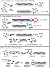
Figure 4. Tmods’ actin-regulatory activities
(A) Tmods transiently cap actin filament pointed ends, reducing actin association/dissociation rates at pointed ends thereby slowing filament growth and shortening [Weber et al. 1994]. (B) Tmods tightly cap TM-actin filament pointed ends, blocking actin association/dissociation and stabilizing filaments [Weber et al., 1994; Weber et al., 1999]. Upon (rare) Tmod dissociation, ends could be converted from TM-actin pointed ends into actin pointed ends, allowing dynamic actin association/dissociation and filament remodeling [Fischer and Fowler, 2003]. When actin subunits polymerize (or depolymerize) up to the end of a TM rod, ends can be converted back into TM-actin pointed ends, with tight capping by Tmod [Fischer and Fowler, 2003; Weber et al., 1999]. (C) Tmods 2 and 3 can bind and sequester monomers, leading to actin filament depolymerization from uncapped ends [Fischer et al., 2006; Yamashiro et al., 2010]. (D) Tmods 1–3 promote actin nucleation, likely by stabilizing a spontaneously formed actin dimer, followed by recruitment of a third monomer to create an actin nucleus, which then polymerizes via elongation from its free barbed end [Yamashiro et al., 2010].
Figure 5
Figure 5. Cellular phenotypes resulting from Tmod depletion or deletion
(A) Normal RBCs have a symmetric, biconcave shape that deforms and withstands circulatory stresses, but Tmod1-null mouse RBCs are sphero-elliptocytic, with reduced deformability and increased osmotic fragility [Moyer et al., 2010]. Dotted lines in top images indicate planes of cross-sections for the cut-away views of RBCs below. (B) Polarized epithelial cells have tall, cuboidal shapes in confluent monolayers, which collapse into shorter, flattened morphologies upon siRNA depletion of Tmod3 [Weber et al., 2007]. (C) Fiber cells in the eye lens are packed with a near-perfect hexagonal geometry. In the Tmod1-null lens, patches of lens fiber cells are irregularly shaped, and fiber cell packing is disordered [Nowak et al., 2009]. Top, Fiber cell orientation in the whole lens, with the dotted outline indicating the equatorial section plane revealing the fiber cell cross-sections depicted below. (D) Neuroblastoma N2a cells extend several long and branched neurites when stimulated by retinoic acid. shRNA depletion of Tmod2 leads to cells with longer neurites, while shRNA depletion of Tmod1 leads to more neurites per cell with no effect on neurite lengths [Fath et al., 2011]. (E) The SR cisternae in skeletal muscle normally have a narrow, flattened morphology, wrapping around well-aligned myofibrils to efficiently deliver and sequester Ca++ for sarcomere contraction and relaxation, respectively. In Tmod1-null muscle, Tmod3 vacates the SR to cap the thin filament pointed ends (see Fig. 6C), leading to SR swelling and abnormal Ca++ handling. The mechanically weakened SR of Tmod1-null muscle is associated with myofibril misalignment [Gokhin and Fowler, 2011a]. (F) Migrating endothelial cells are polarized with a leading lamellipodium and trailing cell body. siRNA depletion of Tmod3 leads to enhanced rates of cell migration, while Tmod3 overexpression leads to loss of cell polarity, with unproductive protrusions and retractions around the cell circumference [Fischer et al., 2003a].
Figure 6
Figure 6. Tmod associations with actin filament architectures and the mechanisms by which actin filaments are remodeled upon Tmod depletion
(A) Tmod1 stabilizes the short TM-coated actin filaments in the spectrin-actin network of the RBC membrane skeleton. (Left) Tmod1-capped short actin filaments at nodes of a quasi-hexagonal lattice connected by spectrin strands on the cytoplasmic surface of the membrane. (Inset) Adducin caps at barbed ends and Tmod1 caps at pointed ends inhibit actin exchange at filament ends, restricting filament lengths. (Right) In Tmod1-null RBCs, the uncapped actin filament nodes are partially disassembled and have variable lengths, leading to a disrupted spectrin-actin lattice. (Inset) The presence of Tmod3 at 1/5th of wild-type Tmod1 levels is insufficient for capping all actin filaments, likely leading to enhanced actin subunit exchange and filament length redistribution in Tmod1-null RBCs [Moyer et al., 2010]. (B) Tmod1 or Tmod3 caps and stabilizes TM-actin filaments in the membrane skeleton of lens fiber cells or on the lateral membranes of polarized epithelial cells, respectively. Upon elimination of Tmod1 or depletion of Tmod3, TM levels are reduced, actin filaments depolymerize, and the spectrin-actin lattice organization on the membrane is disrupted [Nowak et al., 2009; Weber et al., 2007]. (C) Tmod3 links TM5NM1- and TM4-coated γ-actin filaments to sAnk1.5 in the SR at the M-line, stabilizing SR architecture and function. In Tmod1-null muscle, Tmod3 dissociates from sAnk1.5, and relocates to myofibrils to cap the thin filament pointed ends together with Tmod4. This results in partial disassembly of the Tmod3/sAnk1.5/γ-actin/TM5NM1/TM4 filament complex, translocation of the components towards the Z-line, and destabilization of the SR membrane morphology [Gokhin and Fowler, 2011a].

References

    1. Abdi KM, Bennett V. Adducin promotes micrometer-scale organization of beta2-spectrin in lateral membranes of bronchial epithelial cells. Mol Biol Cell. 2008;19(2):536–545. - PMC - PubMed
    1. Alizadeh A, Clark J, Seeberger T, Hess J, Blankenship T, FitzGerald PG. Targeted deletion of the lens fiber cell-specific intermediate filament protein filensin. Invest Ophthalmol Vis Sci. 2003;44(12):5252–5258. - PubMed
    1. Allen DP, Low PS, Dola A, Maisel H. Band 3 and ankyrin homologues are present in eye lens: evidence for all major erythrocyte membrane components in same non-erythroid cell. Biochem Biophys Res Commun. 1987;149(1):266–275. - PubMed
    1. Almenar-Queralt A, Gregorio CC, Fowler VM. Tropomodulin assembles early in myofibrillogenesis in chick skeletal muscle: evidence that thin filaments rearrange to form striated myofibrils. J Cell Sci. 1999a;112(Pt 8):1111–1123. - PubMed
    1. Almenar-Queralt A, Lee A, Conley CA, Ribas de Pouplana L, Fowler VM. Identification of a novel tropomodulin isoform, skeletal tropomodulin, that caps actin filament pointed ends in fast skeletal muscle. J Biol Chem. 1999b;274(40):28466–28475. - PubMed

Publication types

LinkOut - more resources