. 2012;13(3):2985-3008.
doi: 10.3390/ijms13032985. Epub 2012 Mar 6.

BDMC33, A curcumin derivative suppresses inflammatory responses in macrophage-like cellular system: role of inhibition in NF-κB and MAPK signaling pathways

Affiliations

BDMC33, A curcumin derivative suppresses inflammatory responses in macrophage-like cellular system: role of inhibition in NF-κB and MAPK signaling pathways

Ka-Heng Lee et al. Int J Mol Sci. 2012.

Abstract

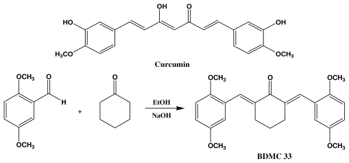
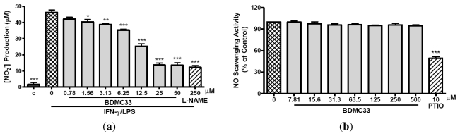
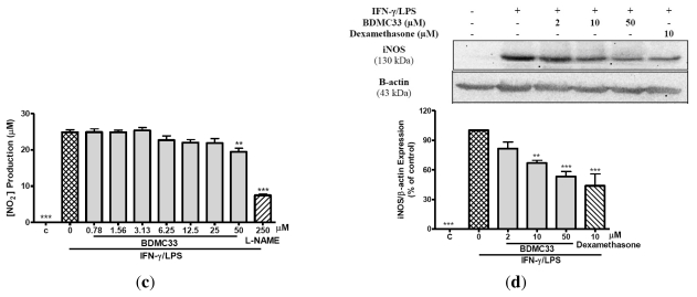
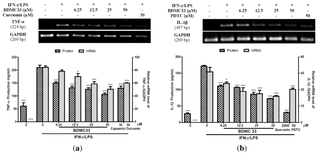
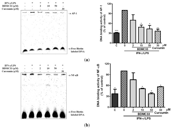
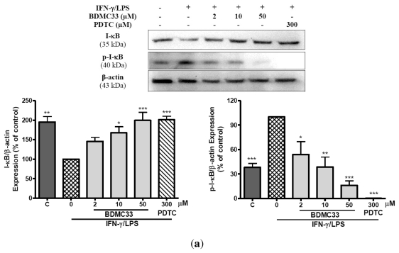
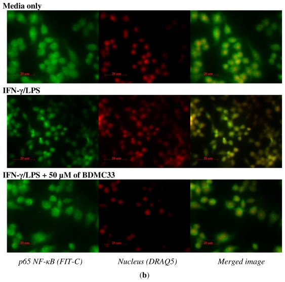
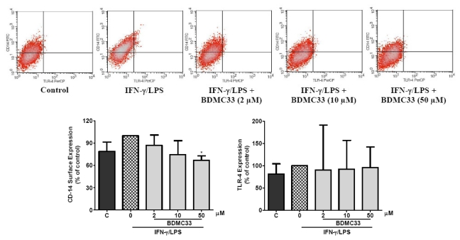
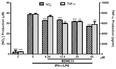
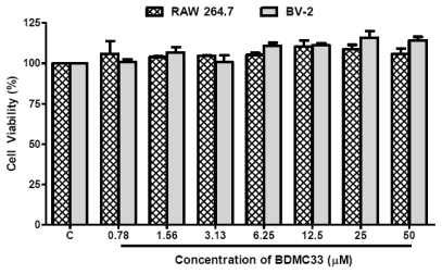
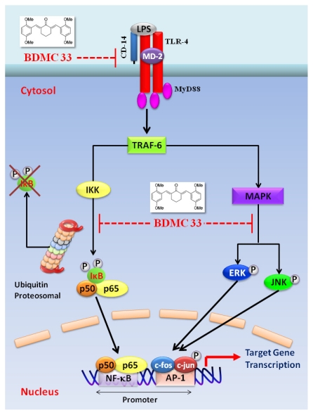
Our preliminary screening has shown that curcumin derivative BDMC33 [2,6-bis(2,5-dimethoxybenzylidene)cyclohexanone] exerted promising nitric oxide inhibitory activity in activated macrophages. However, the molecular basis and mechanism for its pharmacological action is yet to be elucidated. The aim of this study was to investigate the anti-inflammatory properties of BDMC33 and elucidate its underlying mechanism action in macrophage cells. Our current study demonstrated that BDMC33 inhibits the secretion of major pro-inflammatory mediators in stimulated macrophages, and includes NO, TNF-α and IL-1β through interference in both nuclear factor kappaB (NF-κB) and mitogen activator protein kinase (MAPK) signaling cascade in IFN-γ/LPS-stimulated macrophages. Moreover, BDMC33 also interrupted LPS signaling through inhibiting the surface expression of CD-14 accessory molecules. In addition, the inhibitory action of BDMC33 not only restricted the macrophages cell (RAW264.7), but also inhibited the secretion of NO and TNF-α in IFN-γ/LPS-challenged microglial cells (BV-2). The experimental data suggests the inflammatory action of BDMC33 on activated macrophage-like cellular systems, which could be used as a future therapeutic agent in the management of chronic inflammatory diseases.

Keywords: BV-2; MAPK; NF-κB; RAW 264.7; anti-inflammatory; curcumin.


Figures

Figure 1
Figure 1
Chemical structure of curcumin (a) and synthesis of BDMC33 (b).
Figure 2
Figure 2
Effects of BDMC33 on NO production, NO scavenging activity (cell-free system), iNOS activity and iNOS expression in IFN-γ/LPS-induced RAW 264.7 macrophages. (a) Cells were stimulated for 17–20 h with 100 U/mL recombinant murine IFN-γ and 5 μg/mL E. coli LPS and treated with increasing concentrations of BDMC33. The IC50 was calculated at 13.66 ± 0.61 μM. Nitrite level was determined by the Griess reaction after treatment. L-NAME (250 μM) was used as standard iNOS inhibitor for NO inhibition; (b) Percentage of nitrite accumulation produced by sodium nitropruside (SNP) in the presence or absence of BDMC33 was determined by Griess assay. PTIO was used as positive control as a NO scavenger; (c) Cells were treated with IFN-γ/LPS for 12 h prior to treatment with increasing concentrations of BDMC33. L-NAME (250 μM) was used as a standard iNOS inhibitor for NO inhibition. Nitrite level was determined by Griess reaction after treatment; (d) Cells were stimulated for 20 h with combination of IFN-γ/LPS and treated with increasing concentrations of BDMC33. Whole cell lysates were assayed for iNOS expression by using Western blotting. Immunoblotting of β-actin expression was used as loading control. Dexamethasone (DXM) was used as positive control for iNOS expression. The expression level was normalized against IFN-γ/LPS treated group. C, Basal level of nitrite/iNOS expression without IFN-γ/LPS treatment. All values are expressed in mean ± standard error of mean (S.E.M.) of three independent experiments. * P < 0.05, ** P < 0.01, *** P < 0.001 were significantly different from the IFN-γ/LPS-treated control group (second column).
Figure 2
Figure 2
Effects of BDMC33 on NO production, NO scavenging activity (cell-free system), iNOS activity and iNOS expression in IFN-γ/LPS-induced RAW 264.7 macrophages. (a) Cells were stimulated for 17–20 h with 100 U/mL recombinant murine IFN-γ and 5 μg/mL E. coli LPS and treated with increasing concentrations of BDMC33. The IC50 was calculated at 13.66 ± 0.61 μM. Nitrite level was determined by the Griess reaction after treatment. L-NAME (250 μM) was used as standard iNOS inhibitor for NO inhibition; (b) Percentage of nitrite accumulation produced by sodium nitropruside (SNP) in the presence or absence of BDMC33 was determined by Griess assay. PTIO was used as positive control as a NO scavenger; (c) Cells were treated with IFN-γ/LPS for 12 h prior to treatment with increasing concentrations of BDMC33. L-NAME (250 μM) was used as a standard iNOS inhibitor for NO inhibition. Nitrite level was determined by Griess reaction after treatment; (d) Cells were stimulated for 20 h with combination of IFN-γ/LPS and treated with increasing concentrations of BDMC33. Whole cell lysates were assayed for iNOS expression by using Western blotting. Immunoblotting of β-actin expression was used as loading control. Dexamethasone (DXM) was used as positive control for iNOS expression. The expression level was normalized against IFN-γ/LPS treated group. C, Basal level of nitrite/iNOS expression without IFN-γ/LPS treatment. All values are expressed in mean ± standard error of mean (S.E.M.) of three independent experiments. * P < 0.05, ** P < 0.01, *** P < 0.001 were significantly different from the IFN-γ/LPS-treated control group (second column).
Figure 3
Figure 3
(a) Effect of BDMC33 on TNF-α production and gene expression in IFN-γ/LPS-induced RAW 264.7. The IC50 was calculated at 9.40 ± 1.67 μM. Capsaisin (50 μM) and curcumin (30 μM) were used as positive drug control for protein and mRNA expression level of TNF-α respectively; (b) Effect of BDMC33 on IL-1β production and gene expression in IFN-γ/LPS-induced RAW 264.7. The IC50 was calculated at 29.66 ± 0.72 μM. Quercetin (2000 μM) and pyrroline dithiocarbamate (PDTC) (30 μM) were used as positive drug control for protein and mRNA expression level of IL-1β respectively. C; Basal protein and mRNA expression level of TNF-α/IL-1β without IFN-γ/LPS treatment. All values are expressed in mean ± S.E.M. of three independent experiments. * P < 0.05; *** P < 0.001, significantly different from IFN-γ/LPS-treated control group.
Figure 4
Figure 4
Effect of BDMC33 on DNA binding activity of transcriptional factors in IFN-γ/LPS-induced RAW 264.7 macrophages. Cells were stimulated with IFN-γ/LPS and treated with increasing concentrations of BDMC33 for 2 h. The nuclear protein was extracted and tested for DNA binding of (a) AP-1 and (b) NF-κB by EMSA. Curcumin (30 μM) was used as positive drug control for DNA binding activity of both NF-κB and AP-1 transcriptional factors. The expression level was normalized against the IFN-γ/LPS treated group. C, Basal DNA binding activity of NF-κB and AP-1 without IFN-γ/LPS treatment. The results are expressed in mean ± S.E.M. of three independent experiments. * P < 0.05, ** P < 0.01, *** P < 0.001 are significantly different from the IFN-γ/LPS -treated control group (second column).
Figure 5
Figure 5
Effects of BDMC33 on I-κB degradation and I-κB phosphorylation as well as NF-κB nuclear translocation in IFN-γ/LPS induced RAW 264.7 macrophages. (a) Cell lysate analyzed by western blotting using anti-phospho-I-κB and anti-I-κB antibodies. Expression of β-actin expression was used as loading control and normalized against IFN-γ/LPS treated group; (b) Cells cultured in chamber slide were stimulated with IFN-γ/LPS and treated with increasing concentrations of BDMC33. The cellular localization of p65 NF-κB (fluorescein isothiocynate (FTIC) stained, green fluorescent) and nucleus region (DRAQ5 stained, red fluorescent) of cells were identified by immunofluorescence microscopy. All values are the mean ± S.E.M. of three independent experiments. * P < 0.05, ** P < 0.01, *** P < 0.001 are significantly different from IFN-γ/LPS-treated control group.
Figure 5
Figure 5
Effects of BDMC33 on I-κB degradation and I-κB phosphorylation as well as NF-κB nuclear translocation in IFN-γ/LPS induced RAW 264.7 macrophages. (a) Cell lysate analyzed by western blotting using anti-phospho-I-κB and anti-I-κB antibodies. Expression of β-actin expression was used as loading control and normalized against IFN-γ/LPS treated group; (b) Cells cultured in chamber slide were stimulated with IFN-γ/LPS and treated with increasing concentrations of BDMC33. The cellular localization of p65 NF-κB (fluorescein isothiocynate (FTIC) stained, green fluorescent) and nucleus region (DRAQ5 stained, red fluorescent) of cells were identified by immunofluorescence microscopy. All values are the mean ± S.E.M. of three independent experiments. * P < 0.05, ** P < 0.01, *** P < 0.001 are significantly different from IFN-γ/LPS-treated control group.
Figure 6
Figure 6
Effects of BDMC33 on MAPKs activation in IFN-γ/LPS-induced RAW 264.7 macrophages. Cells were stimulated with IFN-γ/LPS and treated with increasing concentrations of BDMC33 for 30 min. Whole cell lysates were analyzed with western blotting with specific antibodies. PD98059, SP600125 and SB203580 were used as standard inhibitors for (a) ERK, (b) JNK and (c) p38 activation, respectively. The ratio of immunointensity between the phospho-MAPKs (p-ERK1/2, p-JNK1/2 and p-p38) and total-MAPKs (ERK1/2, JNK1/2 and p38) were calculated from three independent experiments. Expression of β-actin expression was used as loading control and normalized against IFN-γ/LPS treated group. C; Basal level of MAPK expression without IFN-γ/LPS treatment. The results are expressed in mean ± S.E.M. of three independent experiments. * P < 0.05, ** P < 0.01, *** P < 0.001, significantly different from IFN-γ/LPS-treated control group (second column).
Figure 7
Figure 7
Effects of BDMC33 on IFN-γ/LPS-treated surface expression of TLR-4 and CD-14 accessory molecules in RAW 264.7 macrophages. Cells were stimulated with IFN-γ/LPS and treated with increasing concentrations of BDMC33 for 16 h. The cells were then collected and stained with FITC-conjugated anti-mouse CD-14 and PerCP-conjugated anti-mouse TLR-4 and acquired on FACSCalibur flow cytometer. The expression level was normalized against a IFN-γ/LPS-treated group. C; Basal level of CD-14 or TLR-4 without IFN-γ/LPS treatment. The results are expressed in mean ± S.E.M. of three independent experiments. * P < 0.05, significantly different from IFN-γ/LPS-treated control group (second column).
Figure 8
Figure 8
Effects of BDMC33 on NO and TNF-α production in IFN-γ/LPS-induced BV-2 microglia cells. Cells were stimulated with IFN-γ/LPS and treated with increasing concentrations of BDMC33. Concentrations of NO2 and TNF-α in the media was determined by Griess assay and ELISA, respectively. C, Basal level of NO2/TNF-α without IFN-γ/LPS treatment. All values are the mean ± S.E.M. of three independent experiments. * P < 0.05, ** P < 0.01, *** P < 0.001, significantly different from IFN-γ/LPS-treated control group.
Figure 9
Figure 9
Effects of BDMC33 on cell viability of RAW 264.7 and BV-2 cells. The cells were seeded into a 96-well plate were treated with increasing concentrations of BDMC33 for 24 h and the cell viability determined by MTT assay. C, Basal level of cell viability without IFN-γ/LPS treatment. All values are mean ± S.E.M. of three different independent experiments.
Figure 10
Figure 10
A schematic diagram shows the mechanisms underlying the inhibitory action of BDMC33. TLR-4 stimulation by LPS activates an intracellular signaling cascade that involves the recruitment of MyD88 (myeloid differentiation primary response gene-88) and results in the phosphorylation of TRAF-6 (tumor-necrosis-factor-receptor-associated factor 6), subsequently activate NF-κB and MAPK pathways. The red blunt lines indicate the inhibition by BDMC33.

References

    1. Rodriguez-Vita J., Lawrence T. The resolution of inflammation and cancer. Cytokine Growth Factor Rev. 2010;2:61–65. - PubMed
    1. Schwacha M.G. Macrophages and post-burn immune dysfunction. Burns. 2003;29:1–14. - PubMed
    1. Bowdish D.M.E., Loffredo M.S., Mukhopadhyay S., Mantovani A., Gordon S. Macrophage receptors implicated in the “adaptive” form of innate immunity. Microbes Infect. 2007;9:1680–1687. - PubMed
    1. Chan E.D., Riches D.W.H. IFN-γ + LPS induction of iNOS is modulated by ERK, JNK/SAPK, and p38mapk in a mouse macrophage cell line. Am. J. Physiol. Cell Physiol. 2001;280:441–450. - PubMed
    1. Held T.K., WeiHua X., Yuan L., Kalvakolanu D.V., Cross A.S. Gamma interferon uugments macrophage activation by lipopolysaccharide by two distinct mechanisms, at the signal transduction level and via an autocrine mechanism involving tumor necrosis factor alpha and interleukin-1. Infect. Immun. 1999;67:206–212. - PMC - PubMed

Publication types

MeSH terms