. 2012 May 1;84(9):4153-60.
doi: 10.1021/ac3004055. Epub 2012 Apr 10.

Scanometric microRNA array profiling of prostate cancer markers using spherical nucleic acid-gold nanoparticle conjugates

Affiliations

Scanometric microRNA array profiling of prostate cancer markers using spherical nucleic acid-gold nanoparticle conjugates

Ali H Alhasan et al. Anal Chem. .

Abstract

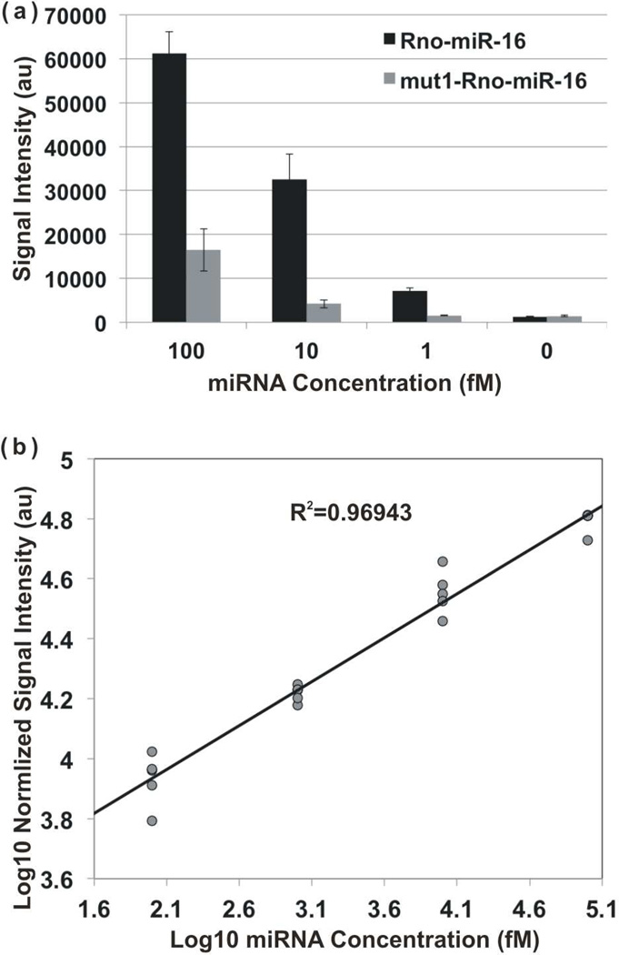
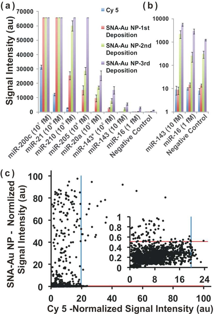
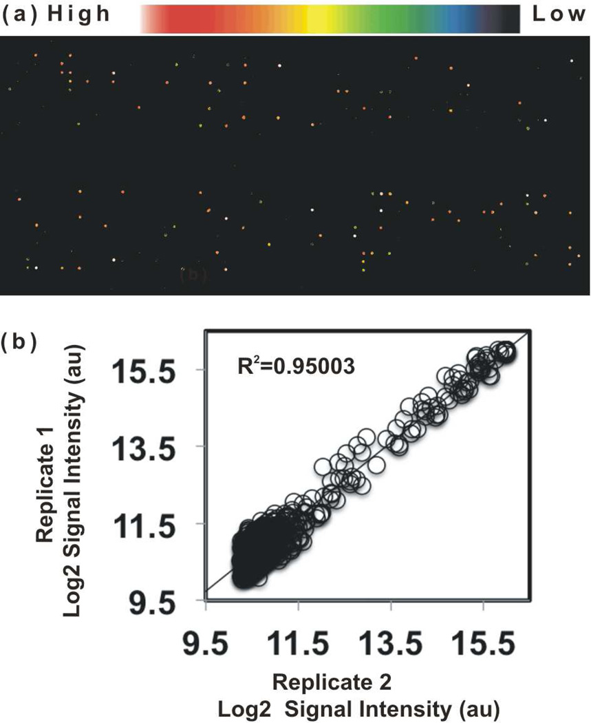
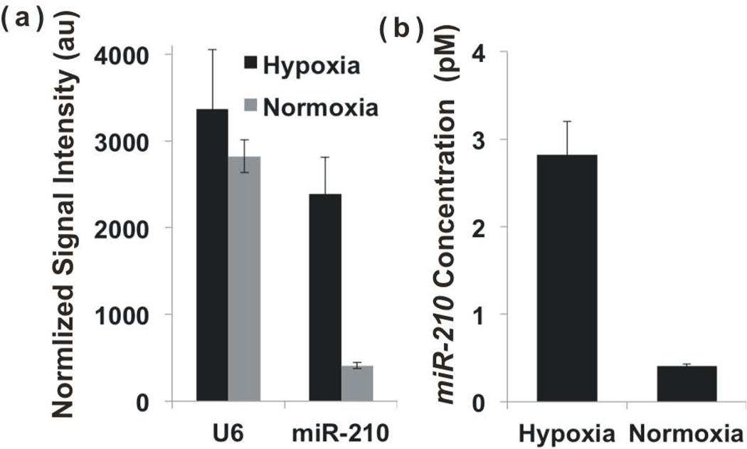
We report the development of a novel Scanometric MicroRNA (Scano-miR) platform for the detection of relatively low abundance miRNAs with high specificity and reproducibility. The Scano-miR system was able to detect 1 fM concentrations of miRNA in serum with single nucleotide mismatch specificity. Indeed, it provides increased sensitivity for miRNA targets compared to molecular fluorophore-based detection systems, where 88% of the low abundance miRNA targets could not be detected under identical conditions. The application of the Scano-miR platform to high density array formats demonstrates its utility for high throughput and multiplexed miRNA profiling from various biological samples. To assess the accuracy of the Scano-miR system, we analyzed the miRNA profiles of samples from men with prostate cancer (CaP), the most common noncutaneous malignancy and the second leading cause of cancer death among American men. The platform exhibits 98.8% accuracy when detecting deregulated miRNAs involved in CaP, which demonstrates its potential utility in profiling and identifying clinical and research biomarkers.


Figures

Figure 1
Figure 1
(a) Synthetic miR-16 was added into denatured human serum at different concentrations and analyzed using the Scano-miR platform. Signal intensities generated from both perfectly matched capture probe sequences (Rno-miR-16) and capture probe with a single nucleotide mismatch (mut1-Rno-miR-16) were plotted. (b) Comparative hybridization of five synthetic miRNAs (miRNA-20a, −143, −143*, −205, and −210) in a Latin square design was performed to complete a five by five matrix (Table S5, see Experimental Sections for experimental details). Plotting signal intensity versus miRNA concentrations generated R2=0.97.
Figure 2
Figure 2
(a) Profile of synthetic miRNAs at different concentrations and (c) Profile of total miRNAs from PC-3 using the Scano-miR assay was plotted and compared to conventional Cy5-based miRNA-array. (b) The low-end detection of (a) is expanded. (c) Inset: scale for the background is expanded.
Figure 3
Figure 3
(a) Detection of human serum miRNAs (from donor 277, Table S6) using the Scano-miR system was performed after hybridizing recovered miRNAs onto a high-density miRNA-array and recorded using the Tecan scanner, where the gray scale image was converted into color using GenePix Pro 6 software (Molecular Devices). Each spot corresponds to a single miRNA and the intensity of the color corresponds to the miRNA concentrations. (b) Biological replicates were analyzed at two different time points using the Scano-miR system. The signal intensity values, calculated using GenePix Pro 6 software, are plotted to calculate the correlation coefficient (R2=0.95). The Log2 of the signal intensities are plotted.
Figure 4
Figure 4
Determining the change in expression level of miR-210 in PC-3 cells in normoxia and hypoxia conditions using (a) the Scano-miR assay and compare the results using (b) qRT-PCR. a) U6 serves as an endogenous loading control. b) Concentrations on y-axis were calculated based on known spiked-in miR-210 concentrations.
Figure 5
Figure 5
A function-gene-miRNA network of 35 deregulated miRNAs was analyzed that target 13 prostate-cancer related genes involved in 5 Gene Ontology terms. A large yellow node in the network represents a single Gene Ontology term, while the small blue nodes stand for genes. Green and red nodes stand for miRNAs with negative and positive logR ratios, respectively. The saturation is proportional to the absolute value of logR ratio.
Scheme 1
Scheme 1
Scheme for the scanometric array-based multiplexed detection of miRNA species (Scano-miR). Isolated miRNAs are enzymatically ligated to a universal linker followed by hybridization onto miRNA microarray. After washing away unbound miRNA species, universal SNA-functionalized gold nanoparticle conjugates (SNA-Au NPs) are subsequently hybridized to detect captured miRNA targets. Next, signal intensity is amplified by depositing gold with gold enhancing solution (1:1 (v:v) mixture of 1 mM HAuCl4 and 10 mM NH2OH) for 5 min and imaged with a Scanner (LS Reloaded, Tecan, Salzburg, Austria).

References

    1. Calin GA, Dumitru CD, Shimizu M, Bichi R, Zupo S, Noch E, Aldler H, Rattan S, Keating M, Rai K, Rassenti L, Kipps T, Negrini M, Bullrich F, Croce CM. Proc. Natl. Acad. Sci. U. S. A. 2002;99:15524. - PMC - PubMed
    1. Nicoloso MS, Spizzo R, Shimizu M, Rossi S, Calin GA. Nat. Rev. Cancer. 2009;9:293. - PubMed
    1. Lee RC, Feinbaum RL, Ambros V. Cell. 1993;75:843. - PubMed
    1. Lim LP, Lau NC, Garrett-Engele P, Grimson A, Schelter JM, Castle J, Bartel DP, Linsley PS, Johnson JM. Nature. 2005;433:769. - PubMed
    1. Lewis BP, Burge CB, Bartel DP. Cell. 2005;120:15. - PubMed

Publication types