. 2012 Dec;88(12):1028-38.
doi: 10.3109/09553002.2012.683511. Epub 2012 May 16.

Induction of lethal bystander effects in human breast cancer cell cultures by DNA-incorporated Iodine-125 depends on phenotype

Affiliations

Induction of lethal bystander effects in human breast cancer cell cultures by DNA-incorporated Iodine-125 depends on phenotype

John M Akudugu et al. Int J Radiat Biol. 2012 Dec.

Abstract

Purpose: This study uses a three-dimensional cell culture model to investigate lethal bystander effects in human breast cancer cell cultures (MCF-7, MDA-MB-231) treated with (125)I-labeled 5-iodo-2 -deoxyuridine ((125)IdU). These breast cancer cell lines respectively form metastatic xenografts in nude mice in an estrogen-dependent and independent manner.

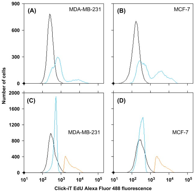
Materials and methods: In the present study, these cells were cultured in loosely-packed three-dimensional architecture in a Cytomatrix™ carbon scaffold. Cultures were pulse-labeled for 3 h with (125)IdU to selectively irradiate a minor fraction of cells, and simultaneously co-pulse-labeled with 0.04 mM 5-ethynyl-2'-deoxyuridine (EdU) to identify the radiolabeled cells using Click-iT(®) EdU and flow cytometry. The cultures were then washed and incubated for 48 h. The cells were then harvested, serially diluted, and seeded for colony formation. Aliquots of cells were subjected to flow cytometry to determine the percentage of cells labeled with (125)IdU/EdU. Additional aliquots were used to determine the mean (125)I activity per labeled cell. The percentage of labeled cells was about 15% and 10% for MCF-7 and MDA cells, respectively. This created irradiation conditions wherein the cross-dose to unlabeled cells was small relative to the self-dose to labeled cells. The surviving fraction relative to EdU-treated controls was measured.

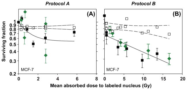
Results: Survival curves indicated significant lethal bystander effect in MCF-7 cells, however, no significant lethal bystander effect was observed in MDA-MB-231 cells.

Conclusions: These studies demonstrate the capacity of (125)IdU to induce lethal bystander effects in human breast cancer cells and suggest that the response depends on phenotype.


Conflict of interest statement

Declaration of interest

The authors report no conflicts of interest. The authors alone are responsible for the content and writing of the paper.

Figures

Figure 1
Figure 1
Representative cellular fluorescence intensity histograms from Protocol B following treatment of MDA-MB-231 cells with (A) 0 mM and (B) 0.04 mM 5-ethynyl-2′-deoxyuridine (EdU); and of MCF-7 cells with (C) 0 mM and (D) 0.04 mM of EdU for 3 h in suspension. Samples corresponding to panels A and C were not exposed to 125IdU, while those corresponding to panels B and D were concomitantly treated with a concentration 0.5 MBq/ml of 125IdU. Gates represent the percentages of cells in each culture that had incorporated EdU (EdU+ or 125IdU/EdU+).
Figure 2
Figure 2
Representative cellular fluorescence intensity histograms following treatment of: (A) MDA-MB-231 cells and (B) MCF-7 cells with 0 mM (black) or with 0.04 mM 5-ethynyl-2′-deoxyuridine (EdU) and 125IdU at a concentration of 1.85 MBq/ml (blue). Cellular fluorescence intensity histograms for cells treated with 0.04 mM EdU and 1.85 MBq/ml of 125IdU, sorted, and then analyzed by flow cytometry into EdU (blue) and EdU+ (brown) populations: (C) MDA-MB-231, (D) MCF-7. Black histograms are for control samples (0 mM EdU).
Figure 3
Figure 3
Surviving fraction of MDA-MB-231 cells as a function of mean absorbed dose to the labeled cell nuclei after treatment with graded amounts of 125IdU using: (A) Protocol A or (B) Protocol B. Solid curves represent least-squares fits of data (filled symbols) to 2-component exponential functions. Solid squares and diamonds represent independent experiments. Dashed lines represent the 95% confidence bands of linear regression fits to the anticipated surviving fraction (1-flabeled, open symbols). Error bars represent SEM of triplicate measurements.
Figure 4
Figure 4
Surviving fraction of MCF-7 cells as a function of mean absorbed dose to the labeled cell nuclei after treatment with varying concentrations of 125IdU using: (A) Protocol A or (B) Protocol B. Solid curves represent least-squares fits of data (filled symbols) to 2-component exponential functions. Solid squares and diamonds represent independent experiments. Dashed lines represent the 95% confidence bands of linear regression fits to the anticipated surviving fraction (1-flabeled, open symbols). Error bars represent SEM of triplicate measurements.
Figure 5
Figure 5
Comparison of measured surviving fractions (colony) with the anticipated surviving fractions for MDA-MB-231 and MCF-7 cells treated with 0.04 mM EdU and varying concentrations of 125IdU, using the Mann-Whitney rank sum test. Only data for absorbed doses wherein the fitted curve for the measured survival was less than or equal to the anticipated survival curve (1-flabeled) were included in the analysis. The horizontal lines within the boxes represent the medians (solid) and means (dashed) of the corresponding data sets, and the error bars represent the standard deviation (n = 7, 8 or 12). A P value of less than 0.05 indicates the difference between the medians of the compared data sets is statistically significant.
Figure 6
Figure 6
Surviving fractions of unlabeled cells as a function of the mean absorbed dose to nuclei of unlabeled cells for: (A) MDA-MB-231 and (B) MCF-7 cells. Solid lines represent two-component exponential fits to the experimental data (filled symbols). Error bars represent propagated standard errors of the mean for triplicate measurements of clonogenic cell survival. These responses are compared to those that would be expected, based on linear quadratic fits of data in the literature, if the unlabeled cells were irradiated acutely with external beams of low linear-energy-transfer ionizing radiation. The dash-dot lines represent published responses for MDA-MB-231: 15 MV photons (α = 0.083 Gy−1, β = 0.0.059 Gy−2, [Jain et al. 2011]) and MCF-7 wild type: 60Co γ-rays (α = 0.59 Gy−1, β = 0.0031 Gy−2, [Lehnert et al. 1990]).

References

    1. Akudugu JM, Neti PVSV, Howell RW. Changes in lognormal shape parameter guide design of patient-specific radiochemotherapy cocktails. Journal of Nuclear Medicine. 2011;52:642–649. - PubMed
    1. Autsavapromporn N, De Toledo SM, Buonanno M, Jay-Gerin JP, Harris AL, Azzam EI. Intercellular communication amplifies stressful effects in high-charge, high-energy (HZE) particle-irradiated human cells. Journal of Radiation Research. 2011;52:408–414. - PMC - PubMed
    1. Azzam EI, de Toledo SM, Gooding T, Little JB. Intercellular communication is involved in the bystander regulation of gene expression in human cells exposed to very low fluences of alpha particles. Radiation Research. 1998;150:497–504. - PubMed
    1. Bishayee A, Rao DV, Howell RW. Evidence for pronounced bystander effects caused by non-uniform distributions of radioactivity using a novel three-dimensional tissue culture model. Radiation Research. 1999;152:88–97. - PMC - PubMed
    1. Bishayee A, Hill HZ, Stein D, Rao DV, Howell RW. Free-radical initiated and gap junction-mediated bystander effect due to nonuniform distribution of incorporated radioactivity in a three-dimensional tissue culture model. Radiation Research. 2001;155:335–344. - PMC - PubMed

Publication types