. 2012 Apr 10:13:54.
doi: 10.1186/1471-2474-13-54.

Rheological and biological properties of a hydrogel support for cells intended for intervertebral disc repair

Affiliations

Rheological and biological properties of a hydrogel support for cells intended for intervertebral disc repair

Karin Benz et al. BMC Musculoskelet Disord. .

Abstract

Background: Cell-based approaches towards restoration of prolapsed or degenerated intervertebral discs are hampered by a lack of measures for safe administration and placement of cell suspensions within a treated disc. In order to overcome these risks, a serum albumin-based hydrogel has been developed that polymerizes after injection and anchors the administered cell suspension within the tissue.

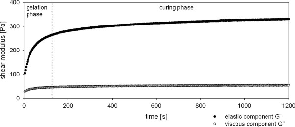
Methods: A hydrogel composed of chemically activated albumin crosslinked by polyethylene glycol spacers was produced. The visco-elastic gel properties were determined by rheological measurement. Human intervertebral disc cells were cultured in vitro and in vivo in the hydrogel and their phenotype was tested by reverse-transcriptase polymerase chain reaction. Matrix production and deposition was monitored by immuno-histology and by biochemical analysis of collagen and glycosaminoglycan deposition. Species specific in situ hybridization was performed to discriminate between cells of human and murine origin in xenotransplants.

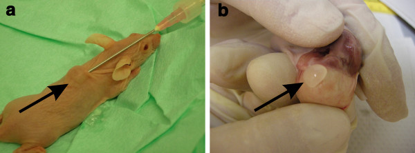
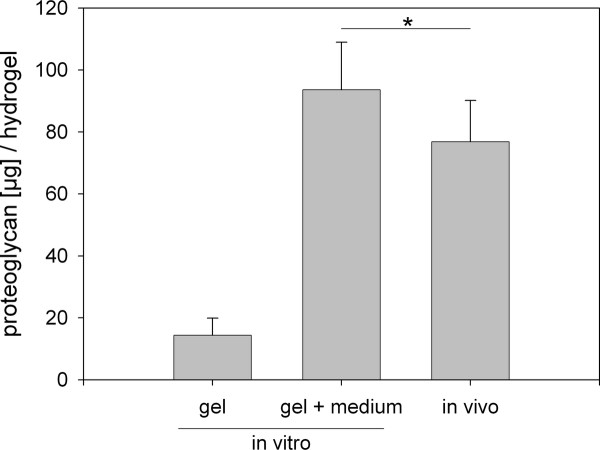
Results: The reproducibility of the gel formation process could be demonstrated. The visco-elastic properties were not influenced by storage of gel components. In vitro and in vivo (subcutaneous implants in mice) evidence is presented for cellular differentiation and matrix deposition within the hydrogel for human intervertebral disc cells even for donor cells that have been expanded in primary monolayer culture, stored in liquid nitrogen and re-activated in secondary monolayer culture. Upon injection into the animals, gels formed spheres that lasted for the duration of the experiments (14 days). The expression of cartilage- and disc-specific mRNAs was maintained in hydrogels in vitro and in vivo, demonstrating the maintenance of a stable specific cellular phenotype, compared to monolayer cells. Significantly higher levels of hyaluronan synthase isozymes-2 and -3 mRNA suggest cell functionalities towards those needed for the support of the regeneration of the intervertebral disc. Moreover, mouse implanted hydrogels accumulated 5 times more glycosaminoglycans and 50 times more collagen than the in vitro cultured gels, the latter instead releasing equivalent quantities of glycosaminoglycans and collagen into the culture medium. Matrix deposition could be specified by immunohistology for collagen types I and II, and aggrecan and was found only in areas where predominantly cells of human origin were detected by species specific in situ hybridization.

Conclusions: The data demonstrate that the hydrogels form stable implants capable to contain a specifically functional cell population within a physiological environment.


Figures

Figure 1
Figure 1
Gelation process of the hydrogel. The graph depicts the gelation process of the hydrogel separated into two phases, the gelation and the curing phase. The two gel components were mixed and injected onto the bottom plate of the rheometer using a dual-chamber applicator syringe. After adjusting the upper plate, the measurement started approximately 30 sec after gel mixture. The experimental conditions of the gel formation were: temperature 37°C, shear strain 0.05%, shear stress 1 Pa, oscillation frequency 1 Hz. Storage modulus G', representing the elastic component of the gel, and loss modulus G", representing the viscous component of the gel, were calculated every third second. The gelation is completed after approximately 2-3 minutes; thereafter the visco-elastic properties of the hydrogel are constant.
Figure 2
Figure 2
mRNA expression profiles of human disc cell preparations cultured in vitro in monolayer or hydrogel. P1 cells were harvested at days 4, 7, and 14 and the expression of collagen type I (COL1), collagen type II (COL2), and aggrecan (ACAN) was analyzed. Expression values of the P1 cultures are expressed relative to the median expression of the cells at the end of the expansion phase (P0) (median because the data are not normally distributed). Boxes represent 25%/75% percentiles, mean values (dotted line) and median values (solid line).
Figure 3
Figure 3
Injection and harvest of cell-loaded hydrogels. A: Injection from dual syringe and 1.3 mm Ø needle. The hydrogel (arrow) polymerizes upon injection and forms a stable subcutaneous protrusion. B: Hydrogel (arrow) at harvest. Note the absence of vascular and connective tissue ingrowth.
Figure 4
Figure 4
Immunohistochemistry staining and species specific genomic in situ hybridization of an implanted hydrogel upon harvest. A: Detection of collagen type I; B: collagen type II; C: aggrecan by immunohistochemistry. The blue dots are the DAPI stained nuclei. Note the even distribution of the nuclei across the samples, except for the edge in A. Species specific in situ hybridization discriminates between cells of human origin (D) and cells of murine origin (E). Mouse cells are predominantly found at the edge of the xenotransplant, human cells are distributed evenly within the transplant.
Figure 5
Figure 5
Glycosaminoglycan production from cells of in vitro maintained and of in vivo implanted hydrogels. Note that most of the glycosaminoglycans (GAG) from the in vitro cultures were found in the culture medium (middle bar). Mean values (n = 6) and standard deviation are shown. In vitro gel: GAG content of the in vitro cultured hydrogel; in vitro gel + medium: GAG content of the in vitro cultured hydrogel plus the GAG released into the combined medium supernatant from two weeks of culture; in vivo: GAG content of the in vivo implanted hydrogel. Statistic: paired t-test comparing the in vitro group (gel + medium) and the in vivo group, * = p < 0.05.
Figure 6
Figure 6
Cumulated collagen production from cells of in vitro maintained and of in vivo implanted hydrogels. Note that most of the collagens from the in vitro cultures were found in the culture medium (middle bar). Mean values (n = 6) and standard deviations are shown. In vitro gel: collagen content of the in vitro cultured hydrogel; in vitro gel + medium: collagen content of the in vitro cultured hydrogel plus the collagen released into the combined medium supernatant from two weeks of culture; in vivo: collagen content of the in vivo implanted hydrogel. Statistic: paired t-test comparing the in vitro group (gel + medium) and the in vivo group, * = p < 0.05.
Figure 7
Figure 7
mRNA expression profiles of the human disc cells in vitro and in vivo. The expression of collagen type I (COL1), collagen type II (COL2), aggrecan (ACAN), hyaluronan-synthase-2 (HAS2), hyaluronan-synthase-3 (HAS3) and SRY (sex determining region Y)-box 9 (SOX9) was analyzed in P2 cells hydrogels cultured for two weeks in vitro or implanted in mice for two weeks and compared to the median expression of the P1 cells (median because not all data were normally distributed). Statistic: One Way repeated Measures Analysis of Variance was performed if the data of all groups were normally distributed, if not, Friedman Repeated Measures Analysis of Variance on Ranks was done. * = p < 0.05. Boxes represent 25%/75% percentiles, mean values (dotted line) and median values (solid line).

References

    1. Meisel HJ, Ganey T, Hutton WC, Libera J, Minkus Y, Alasevic O. Clinical experience in cell-based therapeutics: intervention and outcome. Eur Spine J. 2006;15(Suppl 3):S397–S405. - PMC - PubMed
    1. Meisel HJ, Siodla V, Ganey T, Minkus Y, Hutton WC, Alasevic OJ. Clinical experience in cell-based therapeutics: disc chondrocyte transplantation A treatment for degenerated or damaged intervertebral disc. Biomol Eng. 2007;24(1):5–21. doi: 10.1016/j.bioeng.2006.07.002. - DOI - PubMed
    1. Anderson PA, Rouleau JP. Intervertebral Disc Arthroplasty. Spine. 2004;29(23):2779–2786. doi: 10.1097/01.brs.0000146460.11591.8a. - DOI - PubMed
    1. Boyd L, Carter A. Injectable biomaterials and vertebral endplate treatment for repair and regeneration of the intervertebral disc. Eur Spine J. 2006;15(Suppl 3):S414–S421. - PMC - PubMed
    1. Scholz B, Kinzelmann C, Benz K, Mollenhauer J, Wurst H, Schlosshauer B. Suppression of adverse angiogenesis in an albumin-based hydrogel for articular cartilage and intervertebral disc regeneration. Eur Cell Mater. 2010;20:24–36. discussion 36-27. - PubMed

Publication types

MeSH terms

LinkOut - more resources